File:Gray0986.jpg: Difference between revisions
mNo edit summary |
|||
| (One intermediate revision by the same user not shown) | |||
| Line 2: | Line 2: | ||
Embryo from CRL data appears Week 7 and either [[Carnegie stage 18]] or [[Carnegie stage 19]]. | Embryo from CRL data appears Week 7 and either [[Carnegie stage 18]] or [[Carnegie stage 19]]. | ||
The part of the gut loop on the distal side of the cecal diverticulum increases in diameter and forms the future ascending and transverse portions of the large intestine. Until the fifth month the cecal diverticulum has a uniform caliber, but from this time onward its distal part remains rudimentary and forms the vermiform process, while its proximal part expands to form the cecum. Changes also take place in the shape and position of the stomach. Its dorsal part or greater curvature, to which the dorsal mesogastrium is attached, grows much more rapidly than its ventral part or lesser curvature to which the ventral mesogastrium is fixed. Further, the greater curvature is carried downward and to the left, so that the right surface of the stomach is now directed backward and the left surface forward (Fig. 986), a change in position which explains why the left vagus nerve is found on the front, and the right vagus on the back of the stomach. The dorsal mesogastrium being attached to the greater curvature must necessarily follow its movements, and hence it becomes greatly elongated and drawn lateralward and ventralward from the vertebral column, and, as in the case of the stomach, the right surfaces of both the dorsal and ventral mesogastria are now directed backward, and the left forward. (Gray's Anatomy text) | |||
Latest revision as of 22:36, 30 March 2015
Human Embryo (17mm)
Embryo from CRL data appears Week 7 and either Carnegie stage 18 or Carnegie stage 19.
The part of the gut loop on the distal side of the cecal diverticulum increases in diameter and forms the future ascending and transverse portions of the large intestine. Until the fifth month the cecal diverticulum has a uniform caliber, but from this time onward its distal part remains rudimentary and forms the vermiform process, while its proximal part expands to form the cecum. Changes also take place in the shape and position of the stomach. Its dorsal part or greater curvature, to which the dorsal mesogastrium is attached, grows much more rapidly than its ventral part or lesser curvature to which the ventral mesogastrium is fixed. Further, the greater curvature is carried downward and to the left, so that the right surface of the stomach is now directed backward and the left surface forward (Fig. 986), a change in position which explains why the left vagus nerve is found on the front, and the right vagus on the back of the stomach. The dorsal mesogastrium being attached to the greater curvature must necessarily follow its movements, and hence it becomes greatly elongated and drawn lateralward and ventralward from the vertebral column, and, as in the case of the stomach, the right surfaces of both the dorsal and ventral mesogastria are now directed backward, and the left forward. (Gray's Anatomy text)
- Gray's Images: Development | Lymphatic | Neural | Vision | Hearing | Somatosensory | Integumentary | Respiratory | Gastrointestinal | Urogenital | Endocrine | Surface Anatomy | iBook | Historic Disclaimer
| Historic Disclaimer - information about historic embryology pages |
|---|
 |
| iBook - Gray's Embryology | |
|---|---|

|
|
Reference
Gray H. Anatomy of the human body. (1918) Philadelphia: Lea & Febiger.
Cite this page: Hill, M.A. (2024, May 23) Embryology Gray0986.jpg. Retrieved from https://embryology.med.unsw.edu.au/embryology/index.php/File:Gray0986.jpg
- © Dr Mark Hill 2024, UNSW Embryology ISBN: 978 0 7334 2609 4 - UNSW CRICOS Provider Code No. 00098G
File history
Click on a date/time to view the file as it appeared at that time.
| Date/Time | Thumbnail | Dimensions | User | Comment | |
|---|---|---|---|---|---|
| current | 15:37, 23 August 2009 | 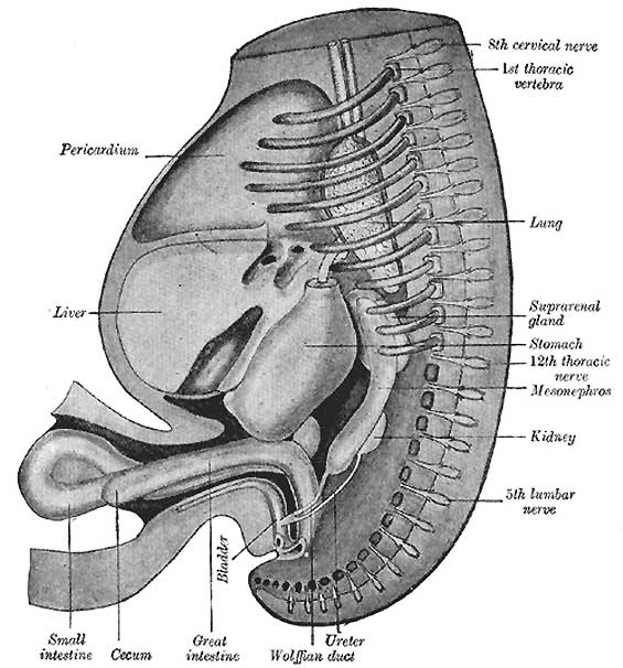 | 565 × 606 (56 KB) | S8600021 (talk | contribs) |
You cannot overwrite this file.
File usage
The following 14 pages use this file:
- 2009 Lecture 9
- 2010 BGD Lecture - Development of the Embryo/Fetus 2
- 2010 BGD Practical 6 - Week 8
- 2010 Lecture 9
- Anatomy of the Human Body by Henry Gray
- BGDA Lecture - Development of the Embryo/Fetus 2
- BGDA Practical 7 - Week 8
- Carnegie stage 18
- Gastrointestinal Tract - Intestine Development
- Gastrointestinal Tract - Mouth Development
- Gastrointestinal Tract - Oesophagus Development
- Gastrointestinal Tract Development
- Lecture - Gastrointestinal Development 2013
- Talk:2010 Lecture 9