File:Gray0931.jpg
Gray0931.jpg (600 × 334 pixels, file size: 53 KB, MIME type: image/jpeg)
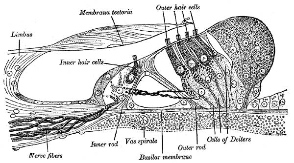
Section through the spiral organ of Corti
Magnified. (G. Retzius.)
The spiral organ of Corti (organon spirale [Corti]; organ of Corti) (Figs. 931, 932) is composed of a series of epithelial structures placed upon the inner part of the basilar membrane. The more central of these structures are two rows of rod-like bodies, the inner and outer rods or pillars of Corti. The bases of the rods are supported on the basilar membrane, those of the inner row at some distance from those of the outer; the two rows incline toward each other and, coming into contact above, enclose between them and the basilar membrane a triangular tunnel, the tunnel of Corti. On the inner side of the inner rods is a single row of hair cells, and on the outer side of the outer rods three or four rows of similar cells, together with certain supporting cells termed the cells of Deiters and Hensen. The free ends of the outer hair cells occupy a series of apertures in a net-like membrane, the reticular membrane, and the entire organ is covered by the tectorial membrane.
- Links: Inner Ear Development
- Gray's Images: Development | Lymphatic | Neural | Vision | Hearing | Somatosensory | Integumentary | Respiratory | Gastrointestinal | Urogenital | Endocrine | Surface Anatomy | iBook | Historic Disclaimer
| Historic Disclaimer - information about historic embryology pages |
|---|
 |
| iBook - Gray's Embryology | |
|---|---|

|
|
Reference
Gray H. Anatomy of the human body. (1918) Philadelphia: Lea & Febiger.
Cite this page: Hill, M.A. (2024, May 21) Embryology Gray0931.jpg. Retrieved from https://embryology.med.unsw.edu.au/embryology/index.php/File:Gray0931.jpg
- © Dr Mark Hill 2024, UNSW Embryology ISBN: 978 0 7334 2609 4 - UNSW CRICOS Provider Code No. 00098G
File history
Click on a date/time to view the file as it appeared at that time.
| Date/Time | Thumbnail | Dimensions | User | Comment | |
|---|---|---|---|---|---|
| current | 00:16, 28 September 2009 | | 600 × 334 (53 KB) | S8600021 (talk | contribs) |
You cannot overwrite this file.
File usage
The following 7 pages use this file: