File:Gray0024.gif
From Embryology
No higher resolution available.
Gray0024.gif (294 × 127 pixels, file size: 5 KB, MIME type: image/gif)
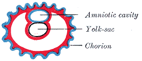
Embryo Week 3
Diagram showing earliest observed stage of human ovum.
- Gray's Images: Development | Lymphatic | Neural | Vision | Hearing | Somatosensory | Integumentary | Respiratory | Gastrointestinal | Urogenital | Endocrine | Surface Anatomy | iBook | Historic Disclaimer
| Historic Disclaimer - information about historic embryology pages |
|---|
 |
| iBook - Gray's Embryology | |
|---|---|

|
|
Reference
Gray H. Anatomy of the human body. (1918) Philadelphia: Lea & Febiger.
Cite this page: Hill, M.A. (2024, May 21) Embryology Gray0024.gif. Retrieved from https://embryology.med.unsw.edu.au/embryology/index.php/File:Gray0024.gif
- © Dr Mark Hill 2024, UNSW Embryology ISBN: 978 0 7334 2609 4 - UNSW CRICOS Provider Code No. 00098G
File history
Click on a date/time to view the file as it appeared at that time.
| Date/Time | Thumbnail | Dimensions | User | Comment | |
|---|---|---|---|---|---|
| current | 14:42, 17 August 2009 | | 294 × 127 (5 KB) | MarkHill (talk | contribs) | Diagram showing earliest observed stage of human ovum. |
You cannot overwrite this file.
File usage
The following page uses this file: