File:Grasshopper heart 01.jpg
Grasshopper_heart_01.jpg (600 × 332 pixels, file size: 58 KB, MIME type: image/jpeg)
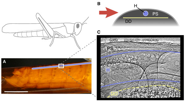
Grasshopper Heart
Flow visualization in the heart of a grasshopper (Schistocerca americana) using synchrotron x-ray phase-contrast imaging.
(A) Side view of the grasshopper abdomen showing the approximate location of the heart (blue) and the relative size of the imaging window (white rectangle, 1.3 × 0.9 mm). The abdomen is encapsulated in an x-ray transparent Kapton tube. Scale bar, 5 mm.
(B), Cross-sectional schematic of the dorsal abdomen showing the relative sizes and locations of the heart (H), dorsal diaphragm (DD), and pericardial sinus (PS). The red arrow indicates the orientation of the x-ray beam.
(C) X-ray video still of a region in the dorsal 3rd abdominal segment in lateral view. Round structures are air bubbles used to visualize patterns of heartbeat and hemolymph flow.
- "With the use of microbubbles as high contrast tracer particles, we directly visualized hemolymph flow in a grasshopper (Schistocerca americana) using synchrotron x-ray phase-contrast imaging. In-vivo intra-heart flow patterns and the relationship between respiratory (tracheae and air sacs) and circulatory (heart) systems were directly observed for the first time."
Original file name: Figure 1. 1472-6793-9-2-1.jpg
Reference
<pubmed>19272159</pubmed>| BMC Physiol.
Lee and Socha BMC Physiology 2009 9:2 doi:10.1186/1472-6793-9-2
© 2009 Lee and Socha; licensee BioMed Central Ltd. This is an Open Access article distributed under the terms of the Creative Commons Attribution License (http://creativecommons.org/licenses/by/2.0), which permits unrestricted use, distribution, and reproduction in any medium, provided the original work is properly cited.
File history
Click on a date/time to view the file as it appeared at that time.
| Date/Time | Thumbnail | Dimensions | User | Comment | |
|---|---|---|---|---|---|
| current | 15:25, 22 November 2010 | | 600 × 332 (58 KB) | S8600021 (talk | contribs) | ==Grasshopper Heart== Flow visualization in the heart of a grasshopper (Schistocerca americana) using synchrotron x-ray phase-contrast imaging. (A) Side view of the grasshopper abdomen showing the approximate location of the heart (blue) and the relativ |
You cannot overwrite this file.
File usage
The following page uses this file: