File:Flecker1932 plate1.jpg

Original file (2,080 × 2,569 pixels, file size: 657 KB, MIME type: image/jpeg)
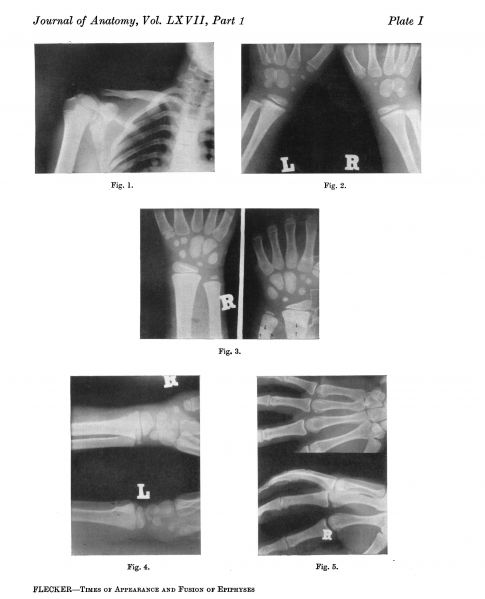
Plate 1
Fig. 1. Boy, age 14 years 7 months. Demonstrating (a) epiphyses at angle of coracoid, (6) tip of coracoid, (c) acromion process, (d) fused head and greater tubercle of humerus.
Fig. 2. Male, age 6 years and 11 months. Multangulum majus well marked on left, but completely absent on the right; lunate just appeared on right but no trace on left.
Fig. 3. Same hands as fig. 4. Note two centres for lunate on left side, one dorsal to other.
Fig. 4. Female, age 6. Note two centres for lunate on left side.
Fig. 5. Sesamoids at heads of all metacarpals. Two sesamoids over fifth.
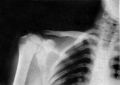
Fig. 1. Boy, age 14 years 7 months. Demonstrating (a) epiphyses at angle of coracoid
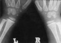
Fig. 2. Male, age 6 years and 11 months. Multangulum majus
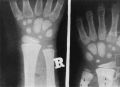
Fig. 3. Same hands as fig. 4.
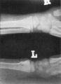
Fig. 4. Female, age 6. Note two centres for lunate on left side.
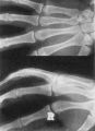
Fig. 5. Sesamoids at heads of all metacarpals.


Reference
Flecker H. (1932). Roentgenographic observations of the times of appearance of epiphyses and their fusion with the diaphyses. (1932) J Anat. 67: 118-164.3 PMID 17104405
Cite this page: Hill, M.A. (2024, April 28) Embryology Flecker1932 plate1.jpg. Retrieved from https://embryology.med.unsw.edu.au/embryology/index.php/File:Flecker1932_plate1.jpg
- © Dr Mark Hill 2024, UNSW Embryology ISBN: 978 0 7334 2609 4 - UNSW CRICOS Provider Code No. 00098G
File history
Click on a date/time to view the file as it appeared at that time.
| Date/Time | Thumbnail | Dimensions | User | Comment | |
|---|---|---|---|---|---|
| current | 22:30, 7 February 2020 | | 2,080 × 2,569 (657 KB) | Z8600021 (talk | contribs) | {{Ref-Flecker1932}} |
You cannot overwrite this file.
File usage
The following page uses this file: