File:Endochondral bone cartoon.jpg

Original file (946 × 513 pixels, file size: 127 KB, MIME type: image/jpeg)
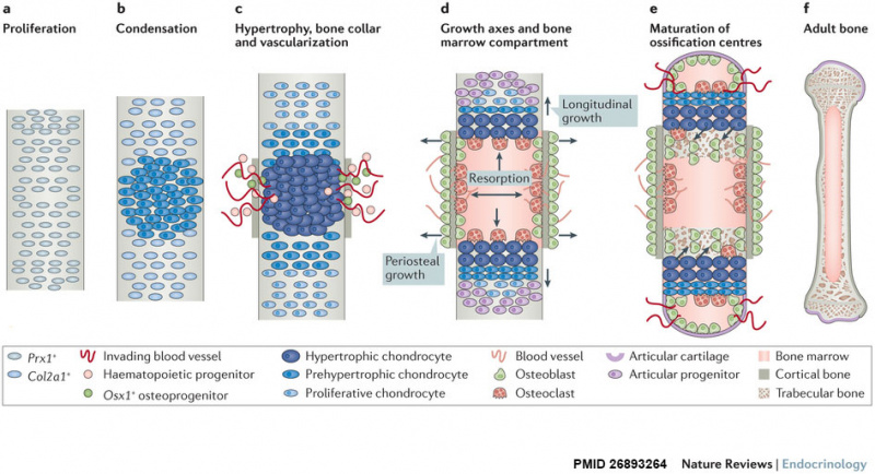
Endochondral Bone Development
Longitudinal views depicting key steps of endochondral bone formation in mouse limbs.
- a - Prx1+ progenitors from lateral plate mesoderm proliferate to populate the emerging limb bud.
- b - Cells nearest the centre undergo mesenchymal condensation, express Col2a1 as they enter a chondrogenic differentiation program, and deposit a cartilage template.
- c to d - Differentiating cells upregulate Col10a1 as they become hypertrophic, which triggers local formation of a bone collar and vascularization of the cartilage template. Invading blood vessels deliver an influx of haematopoietic cells that give rise to osteoclasts which excavate the cartilage template, and Osx1+ osteoblast progenitors and other blood cell types that populate the newly formed marrow cavity.
- d - A longitudinal growth axis is established when vascularization and osteoclast-mediated resorption bisect the presumptive skeletal element, producing two growth plates with opposing directionality. A perpendicular growth axis is driven by periosteal osteoblasts and allows the bone to grow in width.
- e - Within the remodelled cartilage template, bone-forming osteoblasts are derived from Osx1+ cells arriving with the invading vasculature, as well as hypertrophic Col10a1+ chondroctyes that transdifferentiate as they exit the growth plate into the marrow cavity. As bones grow in length and width, a second wave of vascularization forms the secondary ossification centres.
- f - Mature endochondral bone.
Reference
Salazar VS, Gamer LW & Rosen V. (2016). BMP signalling in skeletal development, disease and repair. Nat Rev Endocrinol , 12, 203-21. PMID: 26893264 DOI.
Copyright
Your order details and publisher terms and conditions are available by clicking the link below: http://s100.copyright.com/CustomerAdmin/PLF.jsp?ref=33184fc8-b8d8-4793-8362-2b8e22fcb6c4
Order Details Licensee: Mark A Hill License Date: Sep 11, 2016 License Number: 3946190772930 Publication: Nature Reviews Endocrinology Title: BMP signalling in skeletal development, disease and repair Type Of Use: post on a website
Cite this page: Hill, M.A. (2024, April 28) Embryology Endochondral bone cartoon.jpg. Retrieved from https://embryology.med.unsw.edu.au/embryology/index.php/File:Endochondral_bone_cartoon.jpg
- © Dr Mark Hill 2024, UNSW Embryology ISBN: 978 0 7334 2609 4 - UNSW CRICOS Provider Code No. 00098G
File history
Click on a date/time to view the file as it appeared at that time.
| Date/Time | Thumbnail | Dimensions | User | Comment | |
|---|---|---|---|---|---|
| current | 10:20, 12 September 2016 | | 946 × 513 (127 KB) | Z8600021 (talk | contribs) | ==Endochondral Bone Development== Longitudinal views depicting key steps of endochondral bone formation in mouse limbs. a | Prx1+ progenitors from lateral plate mesoderm proliferate to populate the emerging limb bud. b | Cells nearest the centre under... |
You cannot overwrite this file.
File usage
The following 2 pages use this file: