File:Dandy1910-plate02.jpg

Original file (1,754 × 2,400 pixels, file size: 951 KB, MIME type: image/jpeg)
Plate II
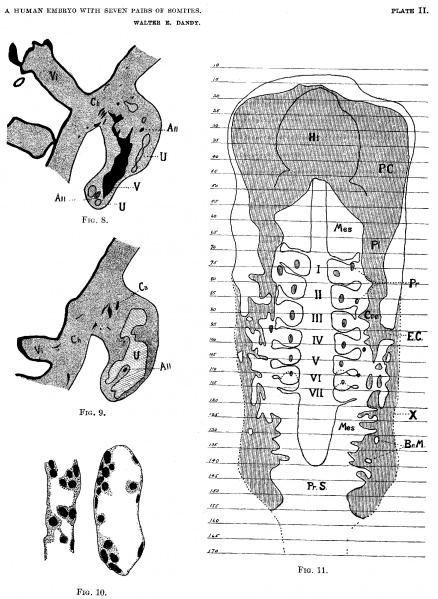
(FIGS. 8-11).
Fig. 8. Section 177, X 25. Outline of section to show size of the sinus formed by union of umbilical veins; V, Umbilical venous sinus (from union of the umbilical veins); All, Allantois, shown in two places, the lower is before division, the upper is terminus of long branch; Ch, Chorion; Vi, Chorionic villus, showing syncytial bud; U, Umbilical artery, shown in two places below, before union to form umbilical arterial sinus, and above, tip of sinus and large branches.
Fig. 9. Section 188, X 25. Outline of section to show size of sinus formed from union of umbilical arteries. U, Umbilical arterial sinus; All, The terminus of the short branch of the allantois; Ca, Capillaries and small branches of sinus in chorion; Ch, Chorionic Membrane; Vi, Chorionic villus, showing capillary entering.
Fig. 10. X 525. Capillaries from chorion, showing apparent formation of blood corpuscles from endothelium of capillaries.
Fig. 11. Reconstruction of coelom, showing relation to somites; Arabic numbers on side represent the numbers of sections; Roman numerals in center represent the paired somites; P.C., Pericardial coelom; Pl, Pleural coelom; Coe, Peritoneal coelom; E.C., External Communication of the coelom: Pr, Pronephros; Ht, Projection of the heart in the pericardial cavity; X. Outer limit of body wall; Mes, Paraxial mesoblat; Br.M., Bridge of mesodermal -tisue extending across the mesodermal slit; Pr.S., Primitive streak.
Online Editor - embryo corresponds to Carnegie stage 10 in Week 4.
| Stage 10 Links: Week 4 | Gastrulation | Lecture | Practical | Image Gallery | Carnegie Embryos | Embryos | Category:Carnegie Stage 10 | Next Stage 11 |
| Historic Papers: 1910 | 1917 | 1926 | 1939 | 1943 | 1957 | 1985 |
| Week: | 1 | 2 | 3 | 4 | 5 | 6 | 7 | 8 |
| Carnegie stage: | 1 2 3 4 | 5 6 | 7 8 9 | 10 11 12 13 | 14 15 | 16 17 | 18 19 | 20 21 22 23 |
| Historic Disclaimer - information about historic embryology pages |
|---|
| Pages where the terms "Historic" (textbooks, papers, people, recommendations) appear on this site, and sections within pages where this disclaimer appears, indicate that the content and scientific understanding are specific to the time of publication. This means that while some scientific descriptions are still accurate, the terminology and interpretation of the developmental mechanisms reflect the understanding at the time of original publication and those of the preceding periods, these terms, interpretations and recommendations may not reflect our current scientific understanding. (More? Embryology History | Historic Embryology Papers) |
- Links: Plate 1 | Plate 2 | Plate 3 | Plate 4 | Plate 5 | Plate 6 | Dandy 1910 | Carnegie stage 10 | Category:Carnegie Stage 10 | Week 4 | Historic Embryology Papers
Reference
Dandy WE. A human embryo with seven pairs of somites measuring about 2 mm in length. (1910) Amer. J Anat. 10: 85-109.
Cite this page: Hill, M.A. (2024, April 27) Embryology Dandy1910-plate02.jpg. Retrieved from https://embryology.med.unsw.edu.au/embryology/index.php/File:Dandy1910-plate02.jpg
- © Dr Mark Hill 2024, UNSW Embryology ISBN: 978 0 7334 2609 4 - UNSW CRICOS Provider Code No. 00098G
File history
Click on a date/time to view the file as it appeared at that time.
| Date/Time | Thumbnail | Dimensions | User | Comment | |
|---|---|---|---|---|---|
| current | 08:48, 18 September 2015 |  | 1,754 × 2,400 (951 KB) | Z8600021 (talk | contribs) | {{Dandy1910 figures}} |
You cannot overwrite this file.
File usage
The following 2 pages use this file:
