File:Cooper1932-fig04.jpg
From Embryology
Size of this preview: 633 × 600 pixels. Other resolution: 900 × 853 pixels.
Original file (900 × 853 pixels, file size: 140 KB, MIME type: image/jpeg)
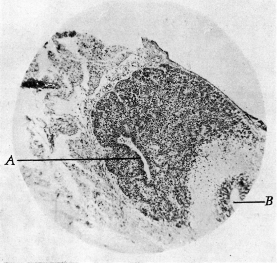
Fig. 4. Sagittal section through pineal region in a foetus of 4 months
x 60. A, cavum pineale. pineele. B, receseus pineale continuous with third ventricle.
| Historic Disclaimer - information about historic embryology pages |
|---|
 |
- Links: pineal
Reference
Cooper ERA. The human pineal gland and pineal cysts. (1932)
Cite this page: Hill, M.A. (2024, May 6) Embryology Cooper1932-fig04.jpg. Retrieved from https://embryology.med.unsw.edu.au/embryology/index.php/File:Cooper1932-fig04.jpg
- © Dr Mark Hill 2024, UNSW Embryology ISBN: 978 0 7334 2609 4 - UNSW CRICOS Provider Code No. 00098G
File history
Click on a date/time to view the file as it appeared at that time.
| Date/Time | Thumbnail | Dimensions | User | Comment | |
|---|---|---|---|---|---|
| current | 17:26, 21 May 2018 | | 900 × 853 (140 KB) | Z8600021 (talk | contribs) | |
| 17:25, 21 May 2018 | 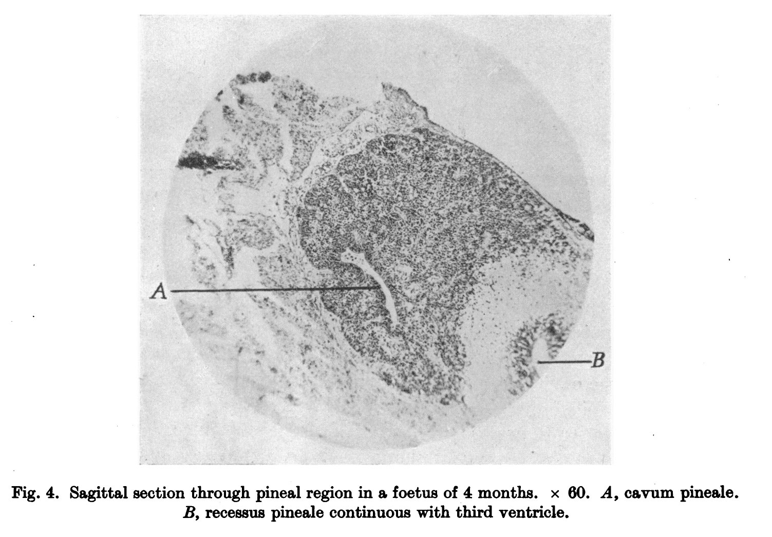 | 1,503 × 1,045 (273 KB) | Z8600021 (talk | contribs) |
You cannot overwrite this file.
File usage
The following 2 pages use this file: