File:Boyden1932 plate02.jpg

Original file (1,000 × 1,698 pixels, file size: 116 KB, MIME type: image/jpeg)
Plate 2
7 Wax reconstruction of blood vessels and kidneys of human embryo shown in plate 1, seen in ventral view. X 55. Note absence of left ureter, fusion of left and right blastemas, the more acute angle at which the left umbilical artery leaves the aorta, the greater size of the left posterior cardinal vein, and the actual contact of blastcmas with the second portion of the posterior cardinal veins (compare Pc./2, fig. 1; 17., fig. 12).
ABBREVIATIONS C’aud.a., caudal artery L.v.com., left vena comitans (compare Il.anast., anastomosis between posterior fig. 1) cardinal veins in iliac region R.isclmr., right ischiatic vein L.med.caud.v., left median caudal vein R.post.card.v., right posterior cardinal vein
Reference
Boyden EA. Congenital absence of the kidney - an interpretation based on a 10-mm human embryo exhibiting unilateral renal agenesis. (1932) Anat. Rec. 52(4):325-349.
Cite this page: Hill, M.A. (2024, May 21) Embryology Boyden1932 plate02.jpg. Retrieved from https://embryology.med.unsw.edu.au/embryology/index.php/File:Boyden1932_plate02.jpg
- © Dr Mark Hill 2024, UNSW Embryology ISBN: 978 0 7334 2609 4 - UNSW CRICOS Provider Code No. 00098G
File history
Click on a date/time to view the file as it appeared at that time.
| Date/Time | Thumbnail | Dimensions | User | Comment | |
|---|---|---|---|---|---|
| current | 10:17, 8 September 2017 | | 1,000 × 1,698 (116 KB) | Z8600021 (talk | contribs) | |
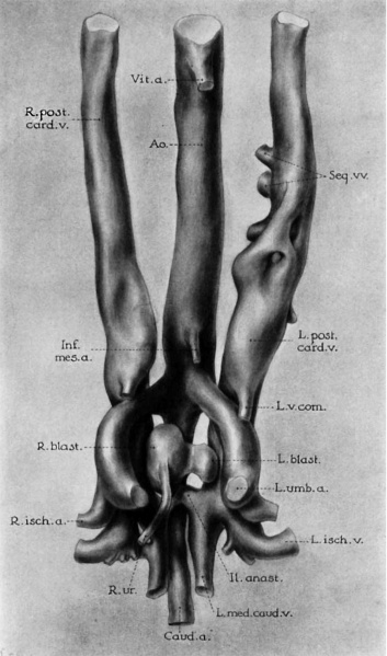
| 10:15, 8 September 2017 | 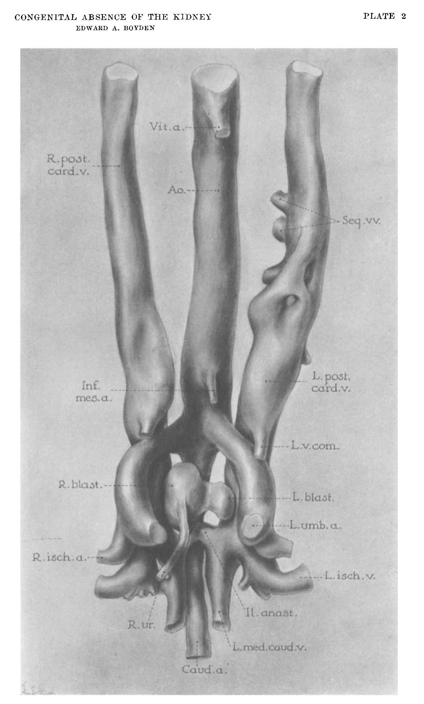 | 1,406 × 2,333 (136 KB) | Z8600021 (talk | contribs) | ===Reference=== {{Ref-Boyden1932}} {{Footer}} |
You cannot overwrite this file.
File usage
The following page uses this file: