File:Boyden1932 plate01.jpg

Original file (1,280 × 907 pixels, file size: 87 KB, MIME type: image/jpeg)
Plate 1
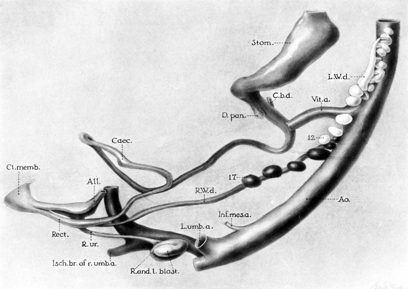
6 Graphic reconstruction of 10 mm human embryo (no. 134 of Minnesota Collection) illustrating arrested development of Wolffian bodies, failure of left Wolffian duct to reach cloaca, and fusion of left blastema with right kidney as latter is entering umbilical crotch. (For relations of veins, see figs. 1 and 7; for crosssections of embryo, see figs. 10 to 12). X 55.
Abbreviations
AIL, allantois Rect., rectum
Ao., aorta R. and L.blast., right and left renal Caec., caecum blastema
Cl.memb., cloacal membrane R.ur., right ureter
C.b.d., common bile duct R.W.d., right Wolffian duct
D.pcm., dorsal pancreas Stom., stomach
Inf.mes.a., inferior mesenteric artery V£t.a., vitelline artery
Isch.br.of 7*.u7nb.a.,rightischiatic artery 1%‘, lowest glomerulus of left Wolfiian L.umb.a., left umbilical artery body
L.W.d., hydronephrotic left Wolffian 1?’, lowest glomerulus of right side duct
Reference
Boyden EA. Congenital absence of the kidney - an interpretation based on a 10-mm human embryo exhibiting unilateral renal agenesis. (1932) Anat. Rec. 52(4):325-349.
Cite this page: Hill, M.A. (2024, May 21) Embryology Boyden1932 plate01.jpg. Retrieved from https://embryology.med.unsw.edu.au/embryology/index.php/File:Boyden1932_plate01.jpg
- © Dr Mark Hill 2024, UNSW Embryology ISBN: 978 0 7334 2609 4 - UNSW CRICOS Provider Code No. 00098G
File history
Click on a date/time to view the file as it appeared at that time.
| Date/Time | Thumbnail | Dimensions | User | Comment | |
|---|---|---|---|---|---|
| current | 10:09, 8 September 2017 | | 1,280 × 907 (87 KB) | Z8600021 (talk | contribs) | |
| 10:08, 8 September 2017 | 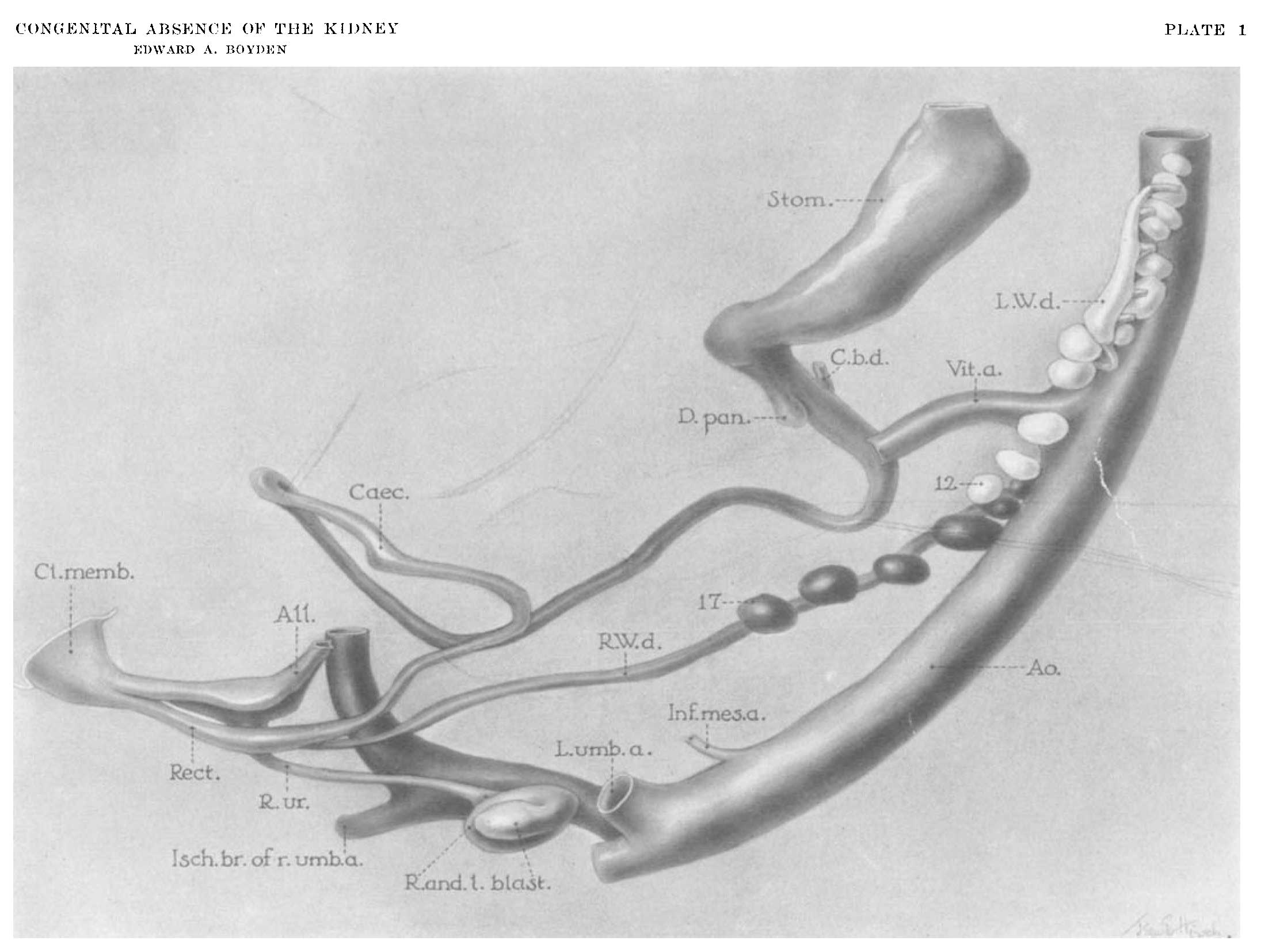 | 2,210 × 1,654 (147 KB) | Z8600021 (talk | contribs) |
You cannot overwrite this file.
File usage
The following page uses this file: