File:Week4.jpg
From Embryology
Size of this preview: 439 × 600 pixels. Other resolution: 521 × 712 pixels.
Original file (521 × 712 pixels, file size: 75 KB, MIME type: image/jpeg)
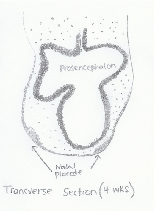
A Transverse section of the forebrain at Week 4 of Embryonic Development. The placodes can be seen as ventrolateral structures arising from the neural crest cells contributed by the prosencephalon. The mesencephalon is also believed to contribute neural crest cells for migration.
Image is self drawn by Student based on the diagram from : <pubmed>15454774</pubmed>
- Note - This image was originally uploaded as part of an undergraduate science student project and may contain inaccuracies in either description or acknowledgements. Students have been advised in writing concerning the reuse of content and may accidentally have misunderstood the original terms of use. If image reuse on this non-commercial educational site infringes your existing copyright, please contact the site editor for immediate removal.
File history
Click on a date/time to view the file as it appeared at that time.
| Date/Time | Thumbnail | Dimensions | User | Comment | |
|---|---|---|---|---|---|
| current | 02:27, 3 October 2012 | | 521 × 712 (75 KB) | Z3331264 (talk | contribs) | Week 4 |
You cannot overwrite this file.
File usage
The following 2 pages use this file: