File:Spleen histology 12.jpg
From Embryology
No higher resolution available.
Spleen_histology_12.jpg (600 × 450 pixels, file size: 93 KB, MIME type: image/jpeg)
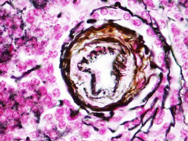
Spleen Histology - Central Arteriole Reticular Fibres
- reticular fibres of the white pulp appear somewhat finer and may be arranged in a series of concentric rings.
- peripheral localisation of the central artery (arteriole) in nodule is quite distinct.
- note the arrangement of reticular fibres around this vessel.
Links: Histology | Histology Stains | Blue Histology images copyright Lutz Slomianka 1998-2009. The literary and artistic works on the original Blue Histology website may be reproduced, adapted, published and distributed for non-commercial purposes. See also the page Histology Stains.
Cite this page: Hill, M.A. (2024, May 23) Embryology Spleen histology 12.jpg. Retrieved from https://embryology.med.unsw.edu.au/embryology/index.php/File:Spleen_histology_12.jpg
- © Dr Mark Hill 2024, UNSW Embryology ISBN: 978 0 7334 2609 4 - UNSW CRICOS Provider Code No. 00098G
File history
Click on a date/time to view the file as it appeared at that time.
| Date/Time | Thumbnail | Dimensions | User | Comment | |
|---|---|---|---|---|---|
| current | 13:43, 26 February 2012 | | 600 × 450 (93 KB) | Z8600021 (talk | contribs) | |
| 13:38, 26 February 2012 | 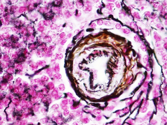 | 665 × 499 (115 KB) | Z8600021 (talk | contribs) | ==Spleen Histology - Central Arteriole Reticular Fibres== * reticular fibres of the white pulp appear somewhat finer and may be arranged in a series of concentric rings. * peripheral localisation of the central artery (arteriole) in nodule is quite dist |
You cannot overwrite this file.
File usage
There are no pages that use this file.