File:Pacinian corpuscle histology 01.jpg
From Embryology
Size of this preview: 750 × 600 pixels.
Original file (800 × 640 pixels, file size: 227 KB, MIME type: image/jpeg)
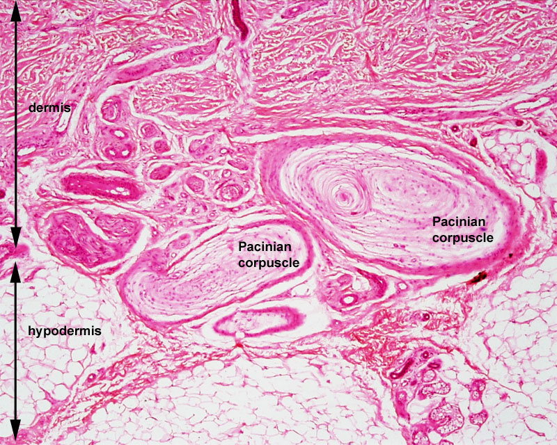
Pacinian Corpuscle Histology
- Pacinian corpuscles are present in the skin and in some mucous membranes.
- mechanoceptors that respond to pressure or any mechanical stimulus deforming the corpuscle.
- generally located deep in the dermis layer near the border with the hypodermis.
Named after Filippo Pacini (1812-1883) an Italian anatomist, published in 1840, and the name "Pacini's corpuscles" was proposed in 1844 by Henle and also by Kölliker.
- Image Links: Labelled Pacinian corpuscle | Unlabelled Pacinian corpuscle | Unlabelled Pacinian corpuscle detail | Meissner's corpuscle image | Touch | Integumentary
Cite this page: Hill, M.A. (2024, June 8) Embryology Pacinian corpuscle histology 01.jpg. Retrieved from https://embryology.med.unsw.edu.au/embryology/index.php/File:Pacinian_corpuscle_histology_01.jpg
- © Dr Mark Hill 2024, UNSW Embryology ISBN: 978 0 7334 2609 4 - UNSW CRICOS Provider Code No. 00098G
File history
Click on a date/time to view the file as it appeared at that time.
| Date/Time | Thumbnail | Dimensions | User | Comment | |
|---|---|---|---|---|---|
| current | 13:56, 26 March 2012 | | 800 × 640 (227 KB) | Z8600021 (talk | contribs) | ==Pacinian Corpuscle Histology== * Pacinian corpuscles are present in the skin and in some mucous membranes. * mechanoceptors that respond to pressure or any mechanical stimulus deforming the corpuscle. |
You cannot overwrite this file.