File:Odgers1939-fig05.jpg
From Embryology
Size of this preview: 476 × 599 pixels. Other resolution: 590 × 743 pixels.
Original file (590 × 743 pixels, file size: 128 KB, MIME type: image/jpeg)
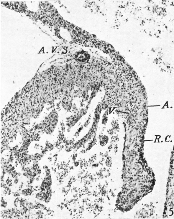
Fig. 5. A section through the right lateral cusp in a 28-5 mm. embryo
( x 60) to show the effect of the invagination which has now occurred at the right A.-V. sulcus, A. V.S. Auricular muscle, A., joins that of the ventricle, V., ventral to the line of attachment of the cusp, so that its base is now composed of auricular muscle, A., and ventricular muscle, 17., with a core of connective tissue continuous with that of the sulcus. The cushion tissue, R.C., extends almost to the tip of the cusp.
File history
Yi efo/eka'e gwa ebo wo le nyangagi wuncin ye kamina wunga tinya nan
| Gwalagizhi | Nyangagi | Dimensions | User | Comment | |
|---|---|---|---|---|---|
| current | 15:01, 15 November 2015 | | 590 × 743 (128 KB) | Z8600021 (talk | contribs) |
You cannot overwrite this file.
File usage
The following 3 pages use this file: