File:Lymph node model dymamics.jpg
Original file (1,000 × 800 pixels, file size: 231 KB, MIME type: image/jpeg)
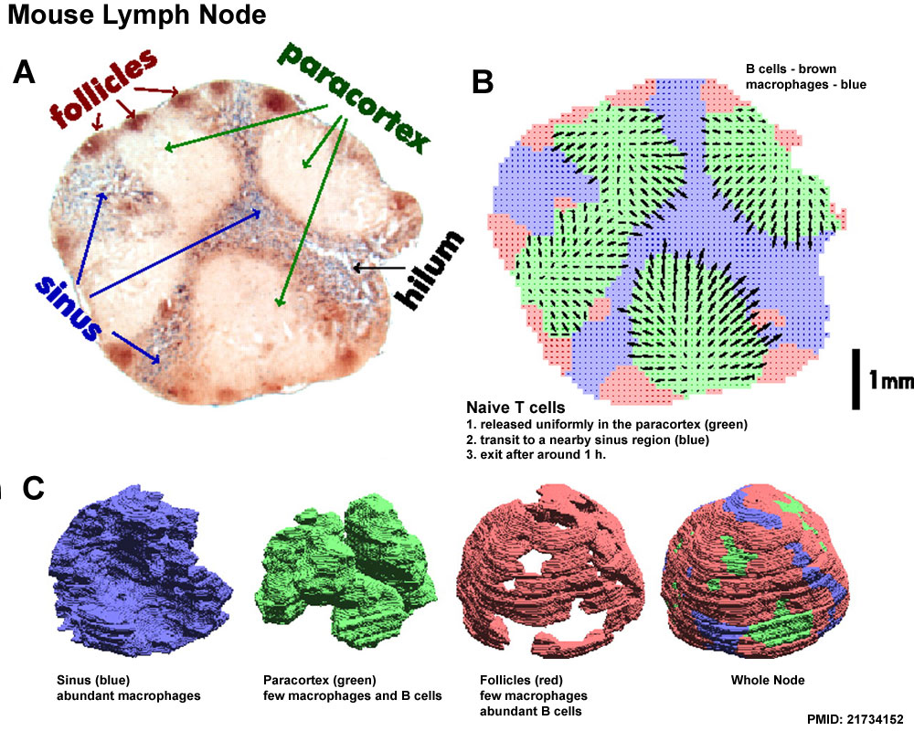
Mouse Lymph Node Dynamics
Modeling the naive T cell lymph node (LN) transit.
A Microtome slice from a mesenteric rat LN with B cells stained in brown and macrophages stained in blue.
B Using image processing algorithms (SI Text), the lymph node volume was divided into three compartments: sinus (blue; abundant macrophages), paracortex (green; few macrophages and B cells), and follicles (red; few macrophages and abundant B cells). 3D renderings of the reconstructed compartments are shown.
C The 3D reconstruction was used to simulate the transit of naive T cells from paracortex to sinus (a 2D projection of the central LN slice is shown).
- The model assumes that naive T cells are released uniformly in the paracortex (green), transit to a nearby sinus region (blue), and then, exit after around 1 h.
- For simulating taxis, a hypothetical chemokine gradient (arrows) pointing from paracortex to sinus was created.
- Arrows indicate direction of the gradient and taxis speed per minute, with arrow lengths magnified 250-fold based on a mean taxis speed of 0.5 μm/min.
Reference
<pubmed>21734152</pubmed>| PMC3145739 | PNAS
Copyright
Proceedings National Academy of Sciences (PNAS) Liberalization of PNAS copyright policy: Noncommercial use freely allowed Note original Author should be contacted for permission to reuse for Educational purposes. See also PNAS Author Rights and Permission FAQs
- Cozzarelli NR, Fulton KR, Sullenberger DM. Liberalization of PNAS copyright policy: noncommercial use freely allowed. Proc Natl Acad Sci U S A. 2004 Aug 24;101(34):12399. PMID15314225 "Our guiding principle is that, while PNAS retains copyright, anyone can make noncommercial use of work in PNAS without asking our permission, provided that the original source is cited."
http://www.pnas.org/content/108/30/12401/F4.expansion.html
Original file name: Fig. 4. Figure has been altered in size and labelling.
File history
Click on a date/time to view the file as it appeared at that time.
| Date/Time | Thumbnail | Dimensions | User | Comment | |
|---|---|---|---|---|---|
| current | 18:15, 22 February 2012 | | 1,000 × 800 (231 KB) | Z8600021 (talk | contribs) | ==Mouse Lymph Node Dynamics== Modeling the naive T cell LN transit. (A) Microtome slice from a mesenteric rat LN with B cells stained in brown and macrophages stained in blue. (B) Using image processing algorithms (SI Text), the lymph node volume was div |
You cannot overwrite this file.
File usage
There are no pages that use this file.