File:Human-spermatozoa.jpg
Human-spermatozoa.jpg (600 × 581 pixels, file size: 19 KB, MIME type: image/jpeg)
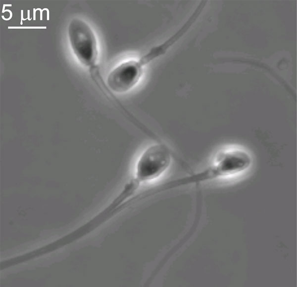
Human Spermatozoa
Human spermatozoa viewed under brightfield phase contrast optics microscopy. Note the general morphology and size of the spermatozoa head and tail.
- Spermatozoa Images: Spermatozoa BF | Spermatozoon BF | Spermatozoon EM | Spermatozoon EM | Historic drawing | Category:Spermatozoa | Spermatozoa Development | Testis Development
Reference
<pubmed>19582168</pubmed>| [1]
Copyright
© 2009 De Blas et al. This is an open-access article distributed under the terms of the Creative Commons Attribution License, which permits unrestricted use, distribution, and reproduction in any medium, provided the original author and source are credited.
Original file name: Figure 1. TRPM8 channels are present in human sperm. (Image from: panel B)
File history
Click on a date/time to view the file as it appeared at that time.
| Date/Time | Thumbnail | Dimensions | User | Comment | |
|---|---|---|---|---|---|
| current | 22:27, 2 May 2010 | | 600 × 581 (19 KB) | S8600021 (talk | contribs) | Human spermatozoa Image from: panel B Figure 1. TRPM8 channels are present in human sperm. A) RT-PCR showing a TRPM8 fragment amplified from human semen. Total RNA was extracted from human semen and cDNA was prepared. Specific oligonucleotides were use |
You cannot overwrite this file.
File usage
The following 10 pages use this file:
- 2010 BGD Lecture - Development of the Embryo/Fetus 1
- 2010 BGD Practical 3 - Gametogenesis
- 2011 Lab 1 - Gametogenesis
- ANAT2341 Lab 1 - Gametogenesis
- BGDA Lecture - Development of the Embryo/Fetus 1
- BGDA Practical 3 - Gametogenesis
- Cell Division - Meiosis
- Lecture - Fertilization
- Spermatozoa Development
- Talk:BGDA Practical 3 - Gametogenesis