File:Hughes1950 fig05.jpg
From Embryology
Size of this preview: 800 × 440 pixels. Other resolution: 1,000 × 550 pixels.
Original file (1,000 × 550 pixels, file size: 49 KB, MIME type: image/jpeg)
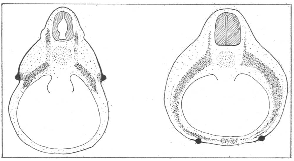
Fig. 5. The epithelial mammary primordium
The epithelial mammary primordium moves ventrally from its original dorsolateral position. This movement is dependent on the anterior growth of the somite.
Figure Links: 5 epithelial mammary primordium | 6 epithelial nipple primordium | Mammary Gland Development
| Historic Disclaimer - information about historic embryology pages |
|---|
 |
Reference
Hughes ES. Development of the mammary gland. (1950) Ann R Coll Surg Engl. 6(2):99-119. PMID 19309885
Cite this page: Hill, M.A. (2024, June 8) Embryology Hughes1950 fig05.jpg. Retrieved from https://embryology.med.unsw.edu.au/embryology/index.php/File:Hughes1950_fig05.jpg
- © Dr Mark Hill 2024, UNSW Embryology ISBN: 978 0 7334 2609 4 - UNSW CRICOS Provider Code No. 00098G
File history
Click on a date/time to view the file as it appeared at that time.
| Date/Time | Thumbnail | Dimensions | User | Comment | |
|---|---|---|---|---|---|
| current | 12:49, 22 March 2018 | | 1,000 × 550 (49 KB) | Z8600021 (talk | contribs) | |
| 12:45, 22 March 2018 | 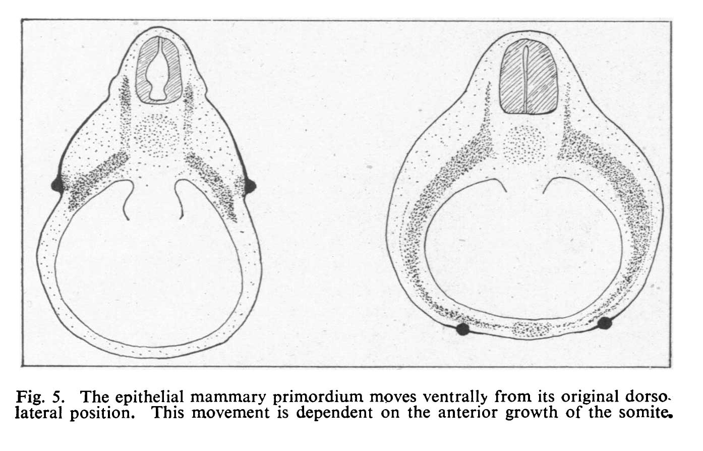 | 1,394 × 894 (101 KB) | Z8600021 (talk | contribs) | {{Hughes1950 figures}} |
You cannot overwrite this file.
File usage
The following page uses this file: