File:Hertig1956 fig82.jpg
Original file (1,000 × 796 pixels, file size: 181 KB, MIME type: image/jpeg)
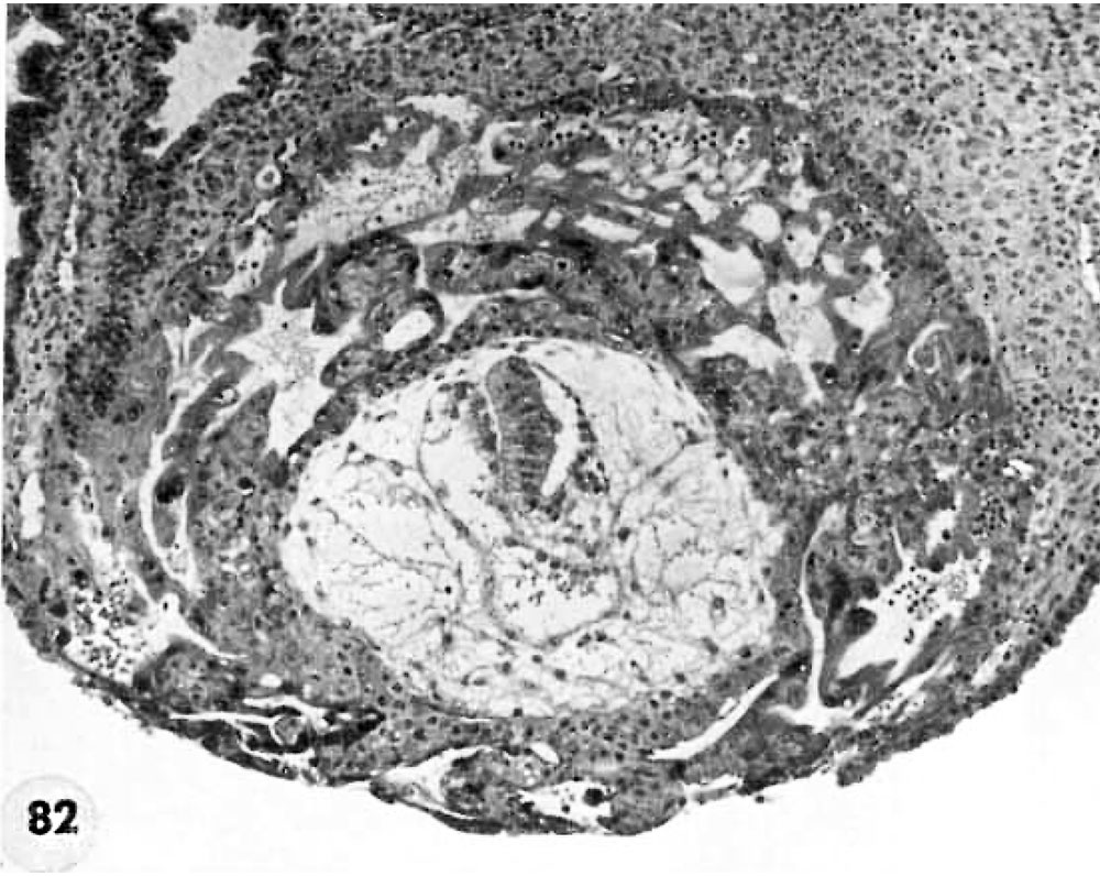
Fig. 82. Abnormal Ova
Two abnormal ova showing malposition of the germ disc and, in one of the specimens, shallow implantation. Whether these ova would ultimately have aborted it is impossible to say.
A medium-power view of the section through the greatest diameter of a 12-day abnormal ovum shown in figures 81 and 83. Note the malposition of the embryonic disc. The normal amniotic development indicates that the malorientation is of recent development since the amnion has been shown to delaminate in situ from adjacent trophoblast at about 7 days of age (Hertig, ’-15). Hence it appears that this normal appearing embryonic. disc has only recently shifted its position due to some unknown factor. It is possible that this maloricntation would have been corrected as the ovum grew and that tile germ disc would have become reoriented. Whether this patient would have aborted ultimately it is impossible to predict, though probably not. It is impossible to say what effect, if any, such an abnormality would have upon the development of the body stalk and the embryo. Carnegie 8299, Section 11-4-5. X 100.
- Figure Links: 1 | 2 | 3 | 4 | 5 | 6 | 7 | 8 | 9-10 | 11-12 | 13-14 | 15-16 | 17 | 18-19 | 20 | 21-22 | 23 | 24-25 | 26-27 | 28-29 | 30-31 | 32-33 | 34 | 35 | 36 | 37 | 38 | 39 | 40 | 41 | 42 | 43 | 44 | 45 | 46 | 47 | 48 | 40 | 49 | 50 | 51 | 52 | 53 | 54 | 55 | 56 | 57 | 58 | 59 | 60 | 61 | 62 | 63 | 64 | 65 | 66 | 67 | 68 | 69 | 70 | 71 | 72 | 73 | 74 | 75 | 76 | 77 | 78 | 79 | 80 | 81 | 82 | 83 | 84 | 85 | 86 | 87 | 88 | 89 | 90 | plate 1 | plate 2 | plate 3 | plate 4 | plate 5 | plate 6 | plate 7 | plate 8 | plate 9 | plate 10 | plate 11 | plate 12 | plate 13 | plate 14 | plate 15 | plate 16 | plate 17 | table 1 | table 1 image | table 2 image | table 3 image | table 4 | table 4 image | table 5 | table 5 image | All figures | 1956 Hertig | Embryology History - Arthur Hertig | John Rock | Historic Papers
Reference
Hertig AT. Rock J. and Adams EC. A description of 34 human ova within the first 17 days of development. (1956) Amer. J Anat., 98:435-493.
Cite this page: Hill, M.A. (2024, May 23) Embryology Hertig1956 fig82.jpg. Retrieved from https://embryology.med.unsw.edu.au/embryology/index.php/File:Hertig1956_fig82.jpg
- © Dr Mark Hill 2024, UNSW Embryology ISBN: 978 0 7334 2609 4 - UNSW CRICOS Provider Code No. 00098G
File history
Click on a date/time to view the file as it appeared at that time.
| Date/Time | Thumbnail | Dimensions | User | Comment | |
|---|---|---|---|---|---|
| current | 14:34, 25 February 2017 | | 1,000 × 796 (181 KB) | Z8600021 (talk | contribs) |
You cannot overwrite this file.
File usage
The following 2 pages use this file: