File:Gray0982b.jpg
Gray0982b.jpg (427 × 393 pixels, file size: 20 KB, MIME type: image/jpeg)
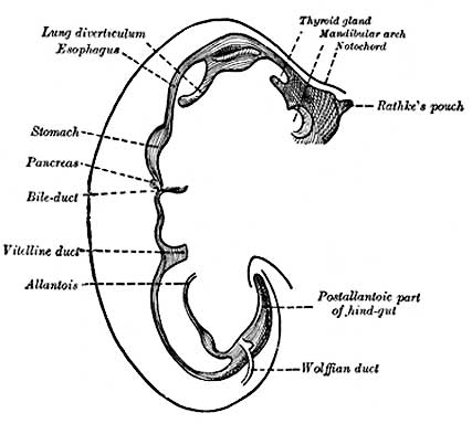
Week 4 - Later Human Digestive Tube
Sketches in profile of two stages in the development of the human digestive tube. (His.) A X 30. B X 20.
About the fourth week a fusiform dilatation, the future stomach, makes its appearance, and beyond this the gut opens freely into the yolk-sac (Fig. 982, A and B). The opening is at first wide, but is gradually narrowed into a tubular stalk, the yolk-stalk or vitelline duct. Between the stomach and the mouth of the yolk-sac the liver diverticulum appears. From the stomach to the rectum the alimentary canal is attached to the notochord by a band of mesoderm, from which the common mesentery of the gut is subsequently developed. The stomach has an additional attachment, viz., to the ventral abdominal wall as far as the umbilicus by the septum transversum. The cephalic portion of the septum takes part in the formation of the diaphragm, while the caudal portion into which the liver grows forms the ventral mesogastrium.
- Gray's Images: Development | Lymphatic | Neural | Vision | Hearing | Somatosensory | Integumentary | Respiratory | Gastrointestinal | Urogenital | Endocrine | Surface Anatomy | iBook | Historic Disclaimer
| Historic Disclaimer - information about historic embryology pages |
|---|
 |
| iBook - Gray's Embryology | |
|---|---|

|
|
Reference
Gray H. Anatomy of the human body. (1918) Philadelphia: Lea & Febiger.
Cite this page: Hill, M.A. (2024, June 5) Embryology Gray0982b.jpg. Retrieved from https://embryology.med.unsw.edu.au/embryology/index.php/File:Gray0982b.jpg
- © Dr Mark Hill 2024, UNSW Embryology ISBN: 978 0 7334 2609 4 - UNSW CRICOS Provider Code No. 00098G
File history
Click on a date/time to view the file as it appeared at that time.
| Date/Time | Thumbnail | Dimensions | User | Comment | |
|---|---|---|---|---|---|
| current | 15:34, 23 August 2009 | | 427 × 393 (20 KB) | S8600021 (talk | contribs) |
You cannot overwrite this file.
File usage
The following 20 pages use this file:
- 2009 Lecture 10
- 2009 Lecture 9
- 2010 Lecture 10
- 2010 Lecture 9
- 2011 Lab 5 - Early Embryo
- ANAT2341 Lab 5 - Early Embryo
- Anatomy of the Human Body by Henry Gray
- BGDB Gastrointestinal - Activity 2
- BGDB Gastrointestinal - Early Embryo
- Draft 2016
- Gastrointestinal Tract - Mouth Development
- Gastrointestinal Tract - Oesophagus Development
- Gastrointestinal Tract Development
- Lecture - Gastrointestinal Development
- Lecture - Gastrointestinal Development 2013
- Lecture - Respiratory Development
- Respiratory System - Upper Respiratory Tract
- Respiratory System Development
- SH Lecture - Respiratory System Development
- Talk:2010 Lecture 9