File:Anatomical diagram of testes.jpg

Original file (647 × 834 pixels, file size: 237 KB, MIME type: image/jpeg)
This is a student drawn image of the testes, epididymis, ductus deferens and spermatic cord.
References
z3417753
Copyright
Beginning six months after publication, I z3417753 grant the public the non-exclusive right to copy, distribute, or display the Work under a Creative Commons Attribution-Noncommercial-Share Alike 3.0 Unported license, as described at http://creativecommons.org/licenses/by-nc-sa/3.0/ and http://creativecommons.org/licenses/by-nc-sa/3.0/legalcode.
--Mark Hill (talk) 10:21, 7 November 2014 (EST) Assessment - Student drawn image, figure relates to project topic contains only copyright and student template. There is no original source provided on which the drawing is based. Figure is clearly drawn. File name is inaccurate is this adult?
- Note - This image was originally uploaded as part of an undergraduate science student project and may contain inaccuracies in either description or acknowledgements. Students have been advised in writing concerning the reuse of content and may accidentally have misunderstood the original terms of use. If image reuse on this non-commercial educational site infringes your existing copyright, please contact the site editor for immediate removal.
File history
Click on a date/time to view the file as it appeared at that time.
| Date/Time | Thumbnail | Dimensions | User | Comment | |
|---|---|---|---|---|---|
| current | 20:29, 20 October 2014 | | 647 × 834 (237 KB) | Z3417753 (talk | contribs) | Reverted to version as of 10:27, 20 October 2014 |
| 20:28, 20 October 2014 |  | 647 × 834 (237 KB) | Z3417753 (talk | contribs) | Reverted to version as of 10:08, 20 October 2014 | |
| 20:27, 20 October 2014 |  | 647 × 834 (237 KB) | Z3417753 (talk | contribs) | Reverted to version as of 10:08, 20 October 2014 | |
| 20:08, 20 October 2014 |  | 647 × 834 (235 KB) | Z3417753 (talk | contribs) | Reverted to version as of 10:05, 20 October 2014 | |
| 20:08, 20 October 2014 | 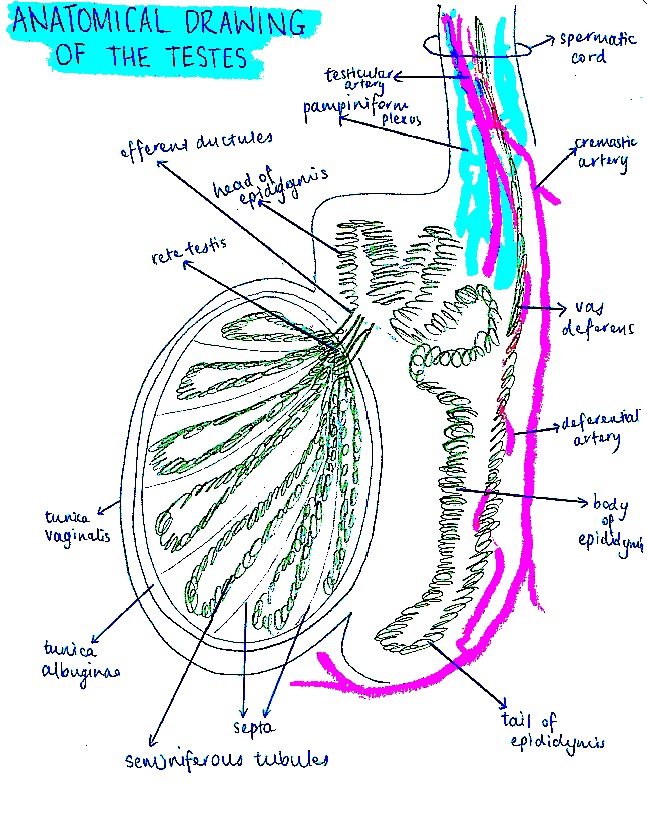 | 647 × 834 (237 KB) | Z3417753 (talk | contribs) | ||
| 20:05, 20 October 2014 | 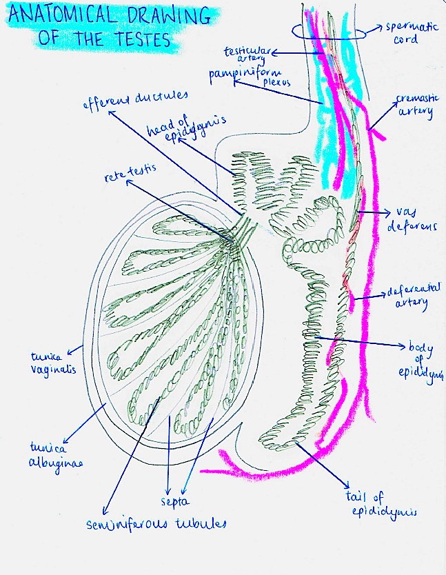 | 647 × 834 (235 KB) | Z3417753 (talk | contribs) | ||
| 19:24, 20 October 2014 |  | 647 × 834 (472 KB) | Z3417753 (talk | contribs) | Student drawn image of the testes, epididymis, ductus deferens and spermatic cord. |
You cannot overwrite this file.
File usage
The following 2 pages use this file: