Dog Development
| Embryology - 19 Apr 2024 |
|---|
| Google Translate - select your language from the list shown below (this will open a new external page) |
|
العربية | català | 中文 | 中國傳統的 | français | Deutsche | עִברִית | हिंदी | bahasa Indonesia | italiano | 日本語 | 한국어 | မြန်မာ | Pilipino | Polskie | português | ਪੰਜਾਬੀ ਦੇ | Română | русский | Español | Swahili | Svensk | ไทย | Türkçe | اردو | ייִדיש | Tiếng Việt These external translations are automated and may not be accurate. (More? About Translations) |
Introduction


The domestic dog (Canis lupus familiaris) has been derived from an ancestoral wolf and now consists of a breed family of more than 300 worldwide, with extensive variations in morphology (size, shape and weight). The modern dog breeds show high phenotypic diversity and are thought to have arisen from this first population bottleneck associated with wolf domestication (7,000–50,000 generations ago) and a second from more recent intensive selection to create the breed (50–100 generations ago).[2]
The average canine gestation period from ovulation to birth (parturition) is approximately 64 days and there have been identified about 400 congenital disorders relating to dog development. Many of these developmental abnormalities are common to human development.
| Dog Links: Introduction | Estrous Cycle | Abnormalities | Category:Dog |
| Historic: 1958 Early Vascular Development | 1961 Foregut Development |
| Animal Development: axolotl | bat | cat | chicken | cow | dog | dolphin | echidna | fly | frog | goat | grasshopper | guinea pig | hamster | horse | kangaroo | koala | lizard | medaka | mouse | opossum | pig | platypus | rabbit | rat | salamander | sea squirt | sea urchin | sheep | worm | zebrafish | life cycles | development timetable | development models | K12 |
Some Recent Findings
|
| More recent papers |
|---|
 This table allows an automated computer search of the external PubMed database using the listed "Search term" text link.
More? References | Discussion Page | Journal Searches | 2019 References | 2020 References Search term: Dog Embryology <pubmed limit=5>Dog Embryology</pubmed> |
Taxon


NCBI Taxonomy Browser Canis lupus familiaris (Genbank common name: dog)
Synonyms: Canis familiaris, Canis domesticus, Canis canis
Chromosomes: 40 (38, X, Y)
Genetic code: Translation table 1 (Standard)
Mitochondrial genetic code: Translation table 2 (Vertebrate Mitochondrial) 16,700 bp
Lineage( full ):cellular organisms; Eukaryota; Fungi/Metazoa group; Metazoa; Eumetazoa; Bilateria; Coelomata; Deuterostomia; Chordata; Craniata; Vertebrata; Gnathostomata; Teleostomi; Euteleostomi; Sarcopterygii; Tetrapoda; Amniota; Mammalia; Theria; Eutheria; Laurasiatheria; Carnivora; Caniformia; Canidae; Canis; Canis lupus

Dog Estrous Cycle
- Proestrus (9 days) - Precedes estrus, estradiol concentration increases as ovarian follicules mature and the uterus enlarges. The vaginal epithelium proliferates accompanied by diapedesis of erythrocytes (most cells in vaginal smear) from uterine capillaries.
- Estrus (9 days) - Accompanied by female mating behaviour, glandular secretions increase, the vaginal epithelium becomes hyperemic, and ovulation occurs. Cycle is influenced mainly by estrogens and the interval between successive estrus cycles is about 7 months.
- Diestrus (70-80 days) - Accompanied by female non-mating behaviour, corpus lutea present and secretes progesterone. Uterine glands undergo hypertrophy and hyperplasia, vaginal secretions and the cervix constricts.
- Anestrus - Anestrus is a prolonged period of sexual rest where the reproductive system is quiescent.
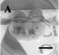
Oocyte Development


Canine geminal vesicle (GV) oocyte.[6]

Dog oocyte development. (A) GV (B) GVBD (C) MI and (D) MII[6]
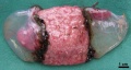
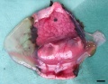
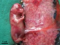
Oocyte to Blastocyst

Canine oocyte to blastocyst (Image: Dr Karine Reynaud).
Development Overview

Days shown below relate to days after ovulation (day 0).
- 48-72 h - oocytes need to complete post-ovulatory maturation to the metaphase II stage in the isthmus of the oviduct[7]
- 2 to 5 days - fertilization
- 14 to 16 days - embryo attaches to uterus
- 22 to 23 days - heartbeat visible
- 62 to 64 days - parturition (birth or whelping)
See also Concannon 2001
Sexual differentiation begins early in the embryonic period prenatally and continues into early postnatal life.
Caudal vena cava development- five theories to origin (right-sided supracardinal, caudal cardinal, sacrocardinal, lateral sympathetic or subcardinal veins).
Carnegie Stages


Gestational age timed from day 0 as the day of the preovulatory serum progesterone rise in the dam, should roughly correlate with fertilisation (+/- 1 day), data[8] *calculated staging only available.
- day 27 - carnegie stage 13.5
- day 28* - carnegie stage 14-15
- day 29* - carnegie stage 15-16
- day 30 - carnegie stage 17
- day 34 - carnegie stage 18.5 – 19
- day 36 - carnegie stage 19.5–20
- day 37 - carnegie stage 20
- Links: Carnegie Stages
E35-38
E35-38
E35-38


Day 28
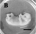
Day 30
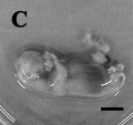
Day 32


Historic Embryology
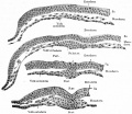
These images are from drawings by Charles Bonnet (1909), later republished in a 1921 textbook of embryology.[9]
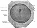
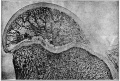
Embryonic disk of dog
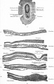
Transverse sections of embryonic disk of dog
Surface view of embryonic disk of dog and transverse sections of same
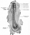
Dorsal view of dog embryo with ten pairs of mesodermal somites
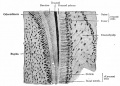
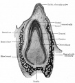
Section through the border of a developing tooth of a new-born puppy
Longitudinal section of a developing tooth of a new-born puppy
Transverse section of dog embryo with ten pairs of primitive segments
Transverse section of a dog embryo with 19 primitive segments
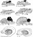
Sagittal sections of different species brains


- Links: Carnegie Stage Comparison

Estrous Cycle
Estrus, also called "in heat" is the time of sexually receptivity and occurs every 17 to 21 days.
- Ovulation occurs 5 to 6 days prior to the first day of diestrus and is indicated by plasma progesterone concentrations higher than 2 ng/mL. (Parturition (birth or whelping) occurs between 62 to 64 days after ovulation).
- Ovulated oocytes diameter[11]
- with the zona pellucida (167.5+/-12.7 microns)
- without zona pellucida (133.9+/-5.3 microns)
- Links: Estrous Cycle
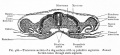
Placenta
Classified as endotheliochorial placentation forming a zonary placenta, which is a complete girdle in dogs.
Three zones:
- girdle zone (endotheliochorial labyrinth)
- hemochorial hemophagous zone (marginal hematoma)
- polar zone (epitheliochorial free)
Trophoblast cell invasion continues after chorioallantois villous penetration and the materno–fetal interface is described as lamellar, with fetal projections interdigitating with maternal septa.
(Data from: Miglino MA, etal., 2006[12] and other sources)
Urogenital System

|

|
| Male Urogenital | Female Urogenital |
Male Gonad
Male sex differentiation is initially mediated by Sry expression then Leydig cell produced testosterone and anti-mullerian hormone (AMH, Mis), also called mullerian-inhibiting substance (MIS) or factor (MIF).
A study using timed pregnancies and male embryo development identified testis differentiation at 36 days gestation. At this time Mullerian duct regression also commenced and was completed by 46 days gestation. Immunohistochemistry also identified Mullerian Inhibitory Substance (MIS) was present during this period in testes and was absent in the undifferentiated testis.[13]
Genital Ridge Sry and Sox9[14]
Testis induction is associated with gonadal Sry and Sox9 expression in mammals, and also with Sox9 expression in vertebrates where Sry is absent. Timing was based upon the equivalent human carnegie staging and expression was measured by quantitative reverse transcription-polymerase chain reaction (qRT-PCR).
- Carnegie Stage 16-18 - Sry expression rose in genital ridge continuously, Sox9 expressed in both male and female genital ridge
- Carnegie Stage 17 - Sox9 expression tenfold greater than in the ovary
- Carnegie Stage 18 - Sry expression maximal
Chromosome 9 Sox 9
Genital Ridge Sf1 and Mis[15]
Mullerian-inhibiting substance - (Mis, Mif) Anti-mullerian hormone (AMH)
Splicing factor 1 - (Sf1) 623 amino acid protein containing a nuclear transport domain, a metal-binding or zinc finger motif, and glutamine- and proline-rich regions.
- Carnegie Stage 15 - Sf1 expression begins in genital ridges
- Carnegie Stage 17 - Sf1 expression pronounced in male and female gonads
- Carnegie Stage 18 - Mis expression only in male gonads
Chromosome 20 Anti-Mullerian hormone
- Links: Sry | Sox9 | Sox 9 Gene | Sf1 | Mis
Spermatogenesis
The cartoons below show nanog expression in the dog during spermatogenesis.[16]

|
Each column represents the combination of different cell types that are present in seminiferous tubules at that specific stage.
Cell types that express NANOG are outlined in red and cell types that do not express NANOG have black and grey symbols. Legend
|
Puberty
| Phase | Rat | Dog (beagle) | Primate (monkey) |
| Neonatal | Birth to postnatal Day 7 | Birth–3 weeks | Birth to 3–4 months |
| Infantile | Postnatal Days 8–21 | 3–5 weeks | Up to 29 months |
| Juvenile/prepubertal | Postnatal Days 22–37 | 5 weeks–6 months | Up to 43 months |
| Pubertal | Postnatal Days 37–38 | 6–8 months | 27–30 months |
Hair Development
Coat variation in the domestic dog is governed by variants in three genes.[18]
- "Coat color and type are essential characteristics of domestic dog breeds. Although the genetic basis of coat color has been well characterized, relatively little is known about the genes influencing coat growth pattern, length, and curl. We performed genome-wide association studies of more than 1000 dogs from 80 domestic breeds to identify genes associated with canine fur phenotypes. Taking advantage of both inter- and intrabreed variability, we identified distinct mutations in three genes, RSPO2, FGF5, and KRT71 (encoding R-spondin-2, fibroblast growth factor-5, and keratin-71, respectively), that together account for most coat phenotypes in purebred dogs in the United States. Thus, an array of varied and seemingly complex phenotypes can be reduced to the combinatorial effects of only a few genes."
Stem Cells
In 2009 a range of canine embryonic stem cell (ESC) lines were developed from preimplantation-stage embryos.[19]
- maintained a normal karyotype and morphology typical of undifferentiated ESCs after multiple in vitro passages and cryopreservation.
- embryoid bodies formed in the absence of a feeder layer in attachment or suspension culture.
- embryoid bodies differentiated into multiple cell types.
- ESCs introduced in vivo formed teratomas containing cell types of all three embryonic germ layers.
Neural
A recent research paper has described a new online digital atlas of the dog brain based upon anatomical and functional magnetic resonance imaging (MRI).[20]
- Links: Database
Abnormalities

There are a number of dog developmental abnormalities that are used as models for human disease.
There are currently 566 abnormality links listed on the Online Mendelian Inheritance in Animals database.
Search OMIA: Canis familiaris
Genital
- Intersex
- Sex reversal - not due to SRY gene translocation to an X chromosome.
Cardiac Defects
- Canine-dilated cardiomyopathy - not associated with canine desmin.[21]
Hip dysplasia
British Veterinary Association and the German Shepherd League scoring scheme
- scoring of nine different radiographic features of each hip
- scale from 0 (ideal) to 6 (worst)
- potential range of subjective scores from 0 to 108.
Other
- Congenital renal disease
- Canine Eclampsia - (puerperal tetany, hypocalcemia) develops mainly in small-breed dogs with large litters.
- Brucellosis - male and female can be carriers of this sexually transmitted disease.
References
- ↑ 1.0 1.1 <pubmed>16311623</pubmed>| PLoS
- ↑ <pubmed>16341006</pubmed>
- ↑ <pubmed>20797342</pubmed>
- ↑ <pubmed>20926804</pubmed>
- ↑ <pubmed>19754575</pubmed>
- ↑ 6.0 6.1 6.2 <pubmed>20565987</pubmed>| Reprod Biol Endocrinol.
- ↑ <pubmed>12620580</pubmed>
- ↑ <pubmed>12840810</pubmed>
- ↑ Bailey, F.R. and Miller, A.M. (1921). Text-Book of Embryology. New York: William Wood and Co. online edition
- ↑ <pubmed>20649949</pubmed>| BMC Genet.
- ↑ <pubmed>17212978</pubmed>
- ↑ <pubmed>16563485</pubmed>
- ↑ <pubmed>1751638</pubmed>
- ↑ <pubmed>12840810</pubmed>
- ↑ <pubmed>15685633</pubmed>
- ↑ <pubmed>20539761</pubmed>| PLoS One.
- ↑ <pubmed>12866705</pubmed>
- ↑ <pubmed>19713490</pubmed>
- ↑ <pubmed>19038794</pubmed>
- ↑ <pubmed>23284904</pubmed>| PMC3527386 | PLoS ONE
- ↑ <pubmed>15475165</pubmed>
Reviews
<pubmed>20797342</pubmed> <pubmed>20537823</pubmed> <pubmed>19942060</pubmed> <pubmed>17560591</pubmed> <pubmed>16564415</pubmed>
Articles
<pubmed>20477984</pubmed> <pubmed>19059739</pubmed> <pubmed>17212978</pubmed> <pubmed>17560591</pubmed> <pubmed>16869883</pubmed> <pubmed>4641196</pubmed> <pubmed>5165787</pubmed> <pubmed>7462099</pubmed>
Online Textbooks
- National Research Council (US) Scientific and Humane Issues in the Use of Random Source Dogs and Cats in Research Committee National Academies Press (US); (2009)
- Proceedings of the November 2003 International Workshop The Development of Science-based Guidelines for Laboratory Animal Care National Academies Press (US); (2004)
Books
- Complete Book of Dog Breeding by DVM Dan Rice
- Practical Dog Breeding: Principles and Practice by William Haynes (1915)
Search Pubmed
Search Pubmed Now: dog development | canine development | Estrous Cycle |
External Links
External Links Notice - The dynamic nature of the internet may mean that some of these listed links may no longer function. If the link no longer works search the web with the link text or name. Links to any external commercial sites are provided for information purposes only and should never be considered an endorsement. UNSW Embryology is provided as an educational resource with no clinical information or commercial affiliation.
- University of Georgia College of Veterinary Medicine The Canine Estrous Cycle
- Comparative Placentation Dog
- A Digital Atlas of the Dog Brain PLoS ONE | Online Data
| Animal Development: axolotl | bat | cat | chicken | cow | dog | dolphin | echidna | fly | frog | goat | grasshopper | guinea pig | hamster | horse | kangaroo | koala | lizard | medaka | mouse | opossum | pig | platypus | rabbit | rat | salamander | sea squirt | sea urchin | sheep | worm | zebrafish | life cycles | development timetable | development models | K12 |
Glossary Links
- Glossary: A | B | C | D | E | F | G | H | I | J | K | L | M | N | O | P | Q | R | S | T | U | V | W | X | Y | Z | Numbers | Symbols | Term Link
Cite this page: Hill, M.A. (2024, April 19) Embryology Dog Development. Retrieved from https://embryology.med.unsw.edu.au/embryology/index.php/Dog_Development
- © Dr Mark Hill 2024, UNSW Embryology ISBN: 978 0 7334 2609 4 - UNSW CRICOS Provider Code No. 00098G