Category:Student Image
From Embryology
Content in this category relates to images added to the site by students.
As shown by the text below:
Note - This image was originally uploaded as part of a student project and may contain inaccuracies in either description or acknowledgements.
--MarkHill 13:50, 4 February 2011 (EST) This category was added in February 2011 and may not appear on images uploaded before this time.
Pages in category 'Student Image'
The following 11 pages are in this category, out of 11 total.
Media in category 'Student Image'
The following 200 files are in this category, out of 650 total.
(previous page) (next page) External genitalia.jpg 783 × 1,633; 576 KB
 Extraocular-muscles-scan.jpg 600 × 532; 76 KB
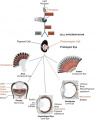
 Eye collage 2.jpg 848 × 417; 112 KB
 Eye diagram bandw.jpg 450 × 369; 42 KB
 Eye evolution.jpg 447 × 563; 106 KB
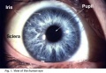 Eye-pupil-sclera-iris.jpg 560 × 389; 52 KB
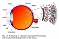 Eye-retina-layers.jpg 726 × 480; 91 KB
 Eyediagramcolour1.JPG 900 × 600; 50 KB
 FASface.jpg 320 × 272; 8 KB
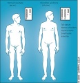
 Female surgeries.jpeg 1,590 × 834; 352 KB
 Ferrier's findings of cerebellum.gif 300 × 167; 42 KB
 Fertilisation of medusa eggs by spermatozoids in vitro in sea water.png 2,877 × 709; 557 KB
 Feta Nail Development - Week 9-10.jpg 367 × 220; 23 KB
 Fetal adrenal gland.png 2,054 × 2,259; 7.7 MB
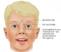
 Fetal alcohol syndrome.jpg 783 × 395; 77 KB
 Fetal Colonic Inury Model Diagram.png 865 × 916; 650 KB
 Fetal Hair Follicle Development - Week 12-14.JPG 573 × 600; 140 KB
 Fetal Hair Follicle Development - Week 13-16.JPG 526 × 600; 127 KB
 Fetal Hair Follicle Development - Week 19-21.JPG 486 × 600; 129 KB
 Fetal Hair Follicle Development - Week 23-28 .JPG 559 × 600; 142 KB
 Fetal Hair Follicle Development - Week 8-12.JPG 571 × 600; 127 KB
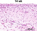 Fetal integumentary histology 14w.jpg 259 × 219; 29 KB
 Fetal integumentary histology Adult.jpg 259 × 217; 30 KB
 Fetal Nail Development - Historic Timeline.jpg 1,007 × 99; 11 KB
 Fetal Nail Development - Week 11-12.jpg 415 × 248; 30 KB
 Fetal Nail Development - Week 13-14 .jpg 437 × 261; 30 KB
 Fetal ovary morphogenesis (mouse).jpg 960 × 681; 93 KB
 Fetal Skeletal Muscle Progenitors.png 1,345 × 1,107; 1.29 MB
 Fetal white blood cell.jpeg 792 × 757; 82 KB
 FGF signalling pathway.jpg 3,507 × 2,550; 1.11 MB
 Fgf signalling.jpg 400 × 219; 46 KB
 FGFR receptor subtype.jpeg 1,092 × 1,334; 130 KB
 Figure 1 Morphological defects in CTCF mutant embryonic hearts.PNG 2,150 × 2,205; 5.9 MB
 Figure 2 - defects of mitochondria in CTCF mutant hearts.PNG 1,807 × 2,400; 2.08 MB
 Figure3testNeuroporeCellShape.png 1,418 × 1,940; 1.01 MB
 Figure5-1.jpg 371 × 450; 48 KB
 FISH for DiGeorge Syndrome.jpg 600 × 470; 31 KB
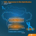 Fluorescent In Situ Hybridisation (FISH).jpg 1,800 × 1,800; 1,016 KB
 Follicular Development.jpeg 661 × 161; 39 KB
 Formation of the choroid and sclera 1.jpg 1,152 × 648; 104 KB
 Formation of the eyelid 1.jpg 1,152 × 648; 100 KB
 Formation of the eyelid 2.jpg 1,152 × 648; 78 KB
 Formation of the lens 1.jpg 1,152 × 648; 102 KB
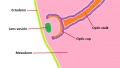 Formation of the lens 2.jpg 1,152 × 648; 94 KB
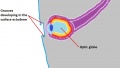
 Formation of the optic cup 1.jpg 1,152 × 648; 47 KB
 Formation of the optic cup 2.jpg 1,152 × 648; 82 KB
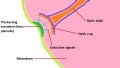
 Formation of the optic nerve and chiasm 1.jpg 1,152 × 648; 59 KB
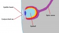
 Formation of the optic vesicle 1.jpg 1,152 × 648; 58 KB
 Formation of the optic vesicle 2.jpg 1,152 × 648; 60 KB
 Formation of the retina 1.jpg 1,152 × 648; 120 KB
 Formation of the retina 2.jpg 1,152 × 648; 121 KB
 Four Models of Lung Branching.jpg 2,537 × 705; 448 KB
 Frataxin Protein.png 800 × 516; 211 KB
 Frog experiments .jpg 580 × 761; 51 KB
 Frog Limb development.jpg 300 × 333; 10 KB
 FSHR Gene.jpg 622 × 207; 33 KB
 Fusion of two pairs of blastomeres inside 4-cell embryos.png 1,247 × 937; 1.39 MB
 Gaba-effects-retina.JPG 879 × 2,084; 240 KB
 Galen-eye1.jpg 1,625 × 1,730; 258 KB
 Gastrula.png 543 × 171; 134 KB
 GEM.jpeg 642 × 472; 83 KB
 GEM.jpg 642 × 472; 83 KB
 Gene expression in the cochlear duct.jpg 600 × 717; 140 KB
 Gene morula.JPG 1,817 × 2,549; 226 KB
 Genes that display strain variation.png 320 × 500; 177 KB
 GIT 2.jpg 2,256 × 1,504; 586 KB
 Glomerular number in Smoke Exposed offspring.png 3,296 × 2,806; 10.85 MB
 Gmb-35-886-g001.jpg 752 × 209; 64 KB
 GnRH receptors (GnRHRs) and spatial expression patterns of gnrhr genes.png 1,941 × 2,790; 6.16 MB
 Granule Cell and Purkinje Cell Migration.png 850 × 673; 399 KB
 Hair Follicle.jpg 2,622 × 2,444; 2.14 MB
 Hand-drawn mammary gland during fetal development.jpg 3,264 × 2,448; 1.42 MB
 Hand-drawn sweat gland development.jpg 3,264 × 2,448; 1.61 MB
 Harlequin Ichthyosis.jpg 520 × 391; 52 KB
 Hatched Blastocyst.jpg 425 × 391; 37 KB
 Hatching .png 377 × 156; 71 KB
 HCC.jpg 600 × 363; 54 KB
 Heart Defects.png 576 × 615; 112 KB
 Heart murmur TOF.png 385 × 343; 172 KB
 Heart Tube Fusion Model of Early Development.png 772 × 1,032; 301 KB
 Heartfibertracttractography.png 2,004 × 1,284; 3.98 MB
 Hedgehog signalling on plane polarity of denticles in Drosophila.png 2,067 × 1,511; 4.12 MB
 Hedgehog.jpg 1,920 × 1,275; 385 KB
 Hemimegalencephaly2.png 1,013 × 746; 510 KB
 Heteroptopia.png 1,010 × 490; 451 KB
 Hh Signalling Pathway.jpg 1,778 × 877; 180 KB
 Hh signalling pathway.jpg 800 × 394; 53 KB
 Hippocampal formation.pdf ; 149 KB
 Histological characteristic during turtle gastrulation.png 2,754 × 1,715; 3.76 MB
 Histology of human embryonic liver at 11 weeks.png 1,988 × 1,642; 4.17 MB
 Histology of Inner Ear.png 592 × 465; 200 KB
 Histopathology of the thymus with hyperplasia.jpg 656 × 385; 256 KB
 Homonculus Sensory and Motor Cortex .png 1,019 × 516; 153 KB
 Horseshoe Kidney.jpg 787 × 787; 205 KB
 Hos.png 1,064 × 1,367; 1 MB
 Human Blastocyst with Decidualized Endometrial Stromal Cells.png 935 × 751; 805 KB
 Human trophoblasts stained with different markers.jpg 784 × 496; 72 KB
 Hydrocele.jpg 343 × 257; 11 KB
 Hypoplastic Left Heart Syndrome (HLHS).png 351 × 464; 254 KB
 Hypospadias.jpg 600 × 325; 53 KB
 Hypothyroidism.jpg 1,492 × 1,634; 565 KB
 ICSI of marmoset oocytes.jpeg 676 × 327; 53 KB
 Illustration of Sox signalling pathway.jpg 837 × 532; 50 KB
 Image.png 588 × 410; 129 KB
 Immunofluorescent FN1 and integrin on blastocytes.gif 100 × 75; 7 KB
 Implantation predictive value of euploid screening results.jpeg 780 × 598; 74 KB
 Infant hearing test.jpg 480 × 640; 82 KB
 Infant with congenital hypothyroidism.jpg 600 × 481; 66 KB
 Integumentary System Fetal Development Timeline.jpg 1,611 × 604; 366 KB
 Internurons migration in cerebral cortex.jpg 510 × 720; 52 KB
 Intro cortex.jpg 960 × 720; 111 KB
 Intro section cortex.jpg 960 × 720; 111 KB
 Introcortex.jpg 960 × 720; 111 KB
 IPSC Formation.gif 539 × 154; 5 KB
 Isthmic Organiser.png 1,235 × 362; 272 KB
 IVF flow chart.png 705 × 189; 50 KB
 Jan Mohr.jpg 1,776 × 2,244; 538 KB
 JNK1.png 441 × 600; 174 KB
 JoubertSyndrome.jpg 633 × 716; 110 KB
 Journal.pbio.0050131.g001.png 1,786 × 2,417; 4.55 MB
 Keibel's model of urogenital organs.jpg 1,478 × 806; 265 KB
 Kidney ascent.jpg 505 × 243; 18 KB
 Kidney position.jpeg 1,630 × 1,204; 600 KB
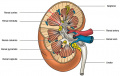 Kidney.jpg 591 × 376; 69 KB
 Klinefelter.jpg 335 × 346; 38 KB
 Knockout mouse process.JPG 1,174 × 578; 79 KB
 Knockoutmouse picture.jpg 772 × 401; 42 KB
 Kollmann691.jpg 729 × 360; 54 KB
 Kollmann692.jpg 603 × 371; 38 KB
 Kollmann693.jpg 693 × 621; 56 KB
 Kollmann694.jpg 686 × 552; 80 KB
 Kollmann697.jpg 731 × 612; 94 KB
 Kollmann698.jpg 689 × 337; 45 KB
 Kollmann699.jpg 722 × 418; 64 KB
 Larvae1.png 610 × 317; 191 KB
 Last try blastula1.png 750 × 163; 181 KB
 LCA fundus and cataracts.jpg 570 × 280; 149 KB
 LCA patient.jpg 464 × 175; 78 KB
 Leber Congential Amaurosis Fundus.jpg 496 × 1,071; 280 KB
 Left and Right Cochlea.jpeg 4,032 × 3,024; 3.45 MB
 Leopard syndrome skin appearance.jpeg 600 × 509; 88 KB
 Leopard syndrome skin appearance.jpg 600 × 509; 88 KB
 LIMB BUD.png 468 × 516; 145 KB
 Lung bud.png 558 × 330; 255 KB
 Lung development overiview.png 717 × 549; 178 KB
 Lung Fgf10 expression cartoon.jpg 1,280 × 821; 96 KB
 Lung Models Normal vs. Diseased.png 600 × 434; 211 KB
 Lung subdivisions cartoon.jpg 600 × 717; 148 KB
 Magnetic Resonance Imaging in an adult patient with tof.JPG 188 × 189; 7 KB
 Malrotation.jpg 483 × 749; 83 KB
 MAS.jpg 640 × 513; 30 KB
 Mature Nail .jpg 554 × 332; 47 KB
 Mechanism of Fertilization.jpg 557 × 298; 41 KB
 Medulloblastoma.jpg 447 × 257; 24 KB
 Melanocyte Pathway.gif 440 × 351; 33 KB
 Melanocytes in Eyes of B6 and ep Mice.jpeg 513 × 634; 112 KB
 Merkel Cell Neurite Complex.JPG 3,264 × 2,448; 2.24 MB
 Merkel Cell Neurite Complexe01.JPG 3,264 × 2,448; 2.18 MB
 Method 1 maternal spindle transfer.png 1,200 × 804; 537 KB
 Method 2 pronuclear transfer.png 1,200 × 752; 592 KB
 Microscopic images of human blastocysts for biopsy.jpeg 673 × 764; 126 KB
 Microtia boy surgery.jpg 655 × 484; 336 KB
 Middle ear ossicles.png 600 × 450; 914 KB
 Midline Prostatic Cyst in Ejaculatory Duct Obstruction.jpeg 786 × 227; 37 KB
 Mip1-expression-in-pitx3.jpg 338 × 600; 63 KB
 Mixed melasma.jpg 600 × 803; 112 KB
 Model of Follicle Development.jpg 600 × 869; 175 KB
 Model of hypercoupled cytoskeleton.png 562 × 600; 101 KB
 Model of the Activities of Cerium Dioxide Nanoparticles.jpeg 472 × 287; 45 KB
 Morphological differences in early mouse embryonic development.png 2,061 × 886; 455 KB
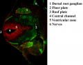
 Morphology of Heart Tube Formation- Student Image .png 681 × 882; 283 KB
 Mouse Kidney Development Cartoon.jpg 506 × 658; 120 KB
 Mouse Neural Crest Contributions to Craniofacial Bones.jpg 600 × 245; 30 KB
 Mouse NT antibody NF Ki67.jpg 1,206 × 945; 607 KB
 Mouse oocytes in vitro.png 3,439 × 2,481; 4.19 MB
 Mouse pronuclei 01.jpg 800 × 801; 51 KB
 Mouse theiler stage 10.JPG 946 × 562; 38 KB
 Mouse theiler stage 11.JPG 689 × 567; 42 KB
 Mouse theiler stage8.JPG 946 × 562; 48 KB
 MRI confirming renal agenesis.jpg 600 × 1,301; 251 KB
 MRI of Goldenhar Syndrome.jpg 357 × 673; 47 KB
 MRI renal agenesis .jpg 600 × 649; 67 KB
 MT transfer.jpg 2,941 × 1,192; 174 KB
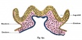
 Mullerian ducts development.jpeg 2,107 × 788; 431 KB
 Mutation of Gene Wt1 Causes Aberrant Gonadal Development.jpeg 600 × 242; 96 KB
 Mutation on GJB2 gene.jpg 306 × 190; 13 KB
 Nadine Dobrovolskaia-Zavadskaia 1948.jpg 240 × 310; 36 KB
 Nail Plate Development - Pinkus .jpg 480 × 270; 19 KB
 Nail Plate Development -Lewis .jpg 692 × 364; 97 KB


.jpg)


.jpg)


_and_spatial_expression_patterns_of_gnrhr_genes.png)


.png)


_image_of_a_transverse_section_of_Sperm_Head_inside_a_cyst_t_the_base_of_a_wild_type_testis_using_Transmission_electron_microscopy.jpeg)


