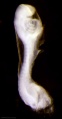
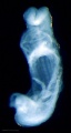
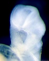
Category:Scanning EM
From Embryology
This page shows UNSW Embryology content related to scanning electron micrographs (SEM) in development.
Note that this may also include some associated bright field images taken before SEM fixation and imaging
- Links: Scanning Electron Microscopy
Pages in category 'Scanning EM'
The following 16 pages are in this category, out of 16 total.
C
Media in category 'Scanning EM'
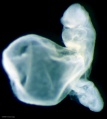
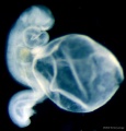
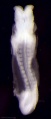
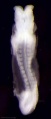
The following 200 files are in this category, out of 426 total.
(previous page) (next page) Anderson2016-fig01.jpg 800 × 800; 93 KB
 Anderson2016-fig02.jpg 800 × 788; 75 KB
 Anderson2016-fig05.jpg 796 × 573; 66 KB
 Cat oocyte zona pellucida 01.jpg 832 × 817; 102 KB
 Cat oocyte zona pellucida 02.jpg 1,000 × 991; 146 KB
 Cat spermatozoa bound to oocyte zona pellucida.jpg 1,000 × 917; 161 KB
 Chicken- PGC grown in vitro 01.jpg 1,174 × 577; 122 KB
 Chicken- PGC grown in vitro 02.jpg 582 × 577; 60 KB
 Chicken- PGC grown in vitro 03.jpg 582 × 577; 61 KB
 Divisions of Early Heart Tube.jpg 1,105 × 978; 85 KB
 Early Heart Tube (Dorsal).jpg 1,282 × 1,124; 111 KB
 Early Heart Tube (Lateral).jpg 1,504 × 972; 110 KB
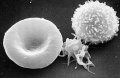 Erythrocyte and lymphocyte SEM01.jpg 800 × 522; 74 KB
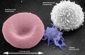 Erythrocyte and lymphocyte SEM02.jpg 800 × 522; 78 KB
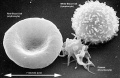 Erythrocyte and lymphocyte SEM03.jpg 800 × 522; 80 KB
 Fetal temporomandibular joint 05.jpg 600 × 392; 71 KB
 Fetal temporomandibular joint 06.jpg 600 × 389; 50 KB
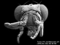
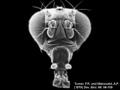
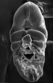
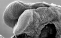
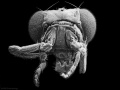 Fly antennapedia head.jpg 900 × 675; 71 KB
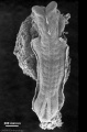
 Fly Hippo-type dorsal view head thorax SEM.jpg 555 × 796; 103 KB
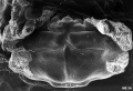
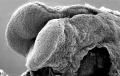
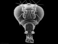 Fly wild-type head.jpg 900 × 675; 54 KB
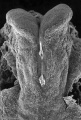
 Fly WT dorsal view head thorax SEM.jpg 555 × 796; 95 KB
 Fly-antennapedia head.jpg 320 × 240; 12 KB
 Fly-wild type head.jpg 320 × 240; 10 KB
 Hamster fused oocyte and spermatozoa.jpg 888 × 405; 98 KB
 Hamster oocyte and spermatozoa.jpg 883 × 836; 266 KB
 Hamster oocyte zona pellucida SEM.jpg 800 × 626; 101 KB
 Heart Looping Sequence (SEMs).jpg 1,928 × 776; 212 KB
 Heart Tube Fusion.jpg 1,551 × 1,139; 125 KB
 Heart Tube Segments.jpg 1,082 × 771; 63 KB
 HeartILP draft HeartTubeDorsal.jpg 1,282 × 1,124; 111 KB
 HeartILP draft hearttubesegments.jpg 1,082 × 771; 63 KB
 HeartILP001.jpg 1,507 × 898; 125 KB
 Human heart SEM1.jpg 1,200 × 330; 47 KB
 Human sperm pathology EM02.jpg 800 × 256; 22 KB
 Human uterine tube ciliated epithelium SEM.jpg 1,200 × 855; 248 KB
 Liver SEM01.jpg 2,000 × 1,333; 350 KB
 Lung epithelium sem11.jpg 1,000 × 999; 240 KB
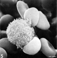
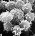
 Lymphocyte rosettes EM01-06.jpg 1,364 × 2,100; 334 KB
 Lymphocyte rosettes EM01.jpg 661 × 665; 58 KB
 Lymphocyte rosettes EM012.jpg 618 × 661; 59 KB
 Lymphocyte rosettes EM02.jpg 661 × 665; 62 KB
 Lymphocyte rosettes EM03.jpg 661 × 665; 51 KB
 Lymphocyte rosettes EM04.jpg 661 × 665; 53 KB
 Lymphocyte rosettes EM05.jpg 661 × 665; 55 KB
 Lymphocyte rosettes EM06.jpg 661 × 665; 66 KB
 ME16 001.jpg 1,740 × 2,500; 557 KB
 ME16 002.jpg 1,037 × 1,500; 272 KB
 ME18 001.jpg 773 × 1,200; 176 KB
 ME34 002.jpg 1,280 × 871; 216 KB
 ME45 002.jpg 2,000 × 1,412; 373 KB
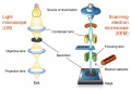 Microscopy LM and SEM cartoon.jpg 720 × 500; 53 KB
 Mouse circumvallate papilla 01.jpg 712 × 955; 114 KB
 Mouse E18.5 cochlea sem01.jpg 902 × 774; 150 KB
 Mouse E18.5 cochlea sem02.jpg 901 × 489; 75 KB
 Mouse E18.5 cochlea sem03.jpg 905 × 1,048; 183 KB
 Mouse E18.5 cochlea sem04.jpg 905 × 535; 85 KB
 Mouse E18.5 cochlea sem05.jpg 807 × 533; 78 KB
 Mouse in vitro follicle 04.jpg 799 × 537; 81 KB
 Mouse in vitro follicle 05.jpg 800 × 639; 82 KB
 Mouse in vitro follicle 06.jpg 800 × 639; 120 KB
 Mouse primitive node cilia.jpg 592 × 981; 130 KB
 Mouse renal podocyte EM01.jpg 1,000 × 1,338; 366 KB
 Mycobacterium-tuberculosis.jpg 320 × 240; 18 KB
 RBC with hereditary persistence of fetal haemoglobin SEM01.jpg 1,200 × 816; 220 KB
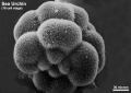
 Sea urchin SEM01.jpg 1,000 × 712; 95 KB
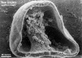
 Sea urchin SEM02.jpg 1,000 × 712; 120 KB
 Sea urchin SEM03.jpg 1,000 × 712; 154 KB
 Sickle cell RBC SEM01.jpg 1,000 × 750; 219 KB
 Stage 9 SEM1.jpg 347 × 450; 42 KB
 Stage10 bf1.jpg 1,000 × 748; 39 KB
 Stage10 bf1a.jpg 800 × 598; 29 KB
 Stage10 bf1b.jpg 600 × 449; 19 KB
 Stage10 bf1c.jpg 400 × 299; 10 KB
 Stage10 bf2.jpg 1,000 × 809; 29 KB
 Stage10 bf2c.jpg 400 × 323; 8 KB
 Stage10 bf3.jpg 836 × 1,000; 35 KB
 Stage10 SEM1.jpg 277 × 450; 28 KB
 Stage10 sem1.jpg 1,000 × 484; 46 KB
 Stage10 sem10.jpg 1,000 × 740; 68 KB
 Stage10 sem10a.jpg 800 × 592; 48 KB
 Stage10 sem10b.jpg 600 × 444; 32 KB
 Stage10 sem10c.jpg 400 × 296; 16 KB
 Stage10 sem11.jpg 1,000 × 636; 76 KB
 Stage10 sem11a.jpg 800 × 509; 59 KB
 Stage10 sem11b.jpg 600 × 382; 38 KB
 Stage10 sem11c.jpg 400 × 255; 20 KB
 Stage10 sem12.jpg 657 × 1,000; 57 KB
 Stage10 sem12a.jpg 526 × 800; 40 KB
 Stage10 sem12b.jpg 395 × 600; 25 KB
 Stage10 sem12c.jpg 263 × 400; 13 KB
 Stage10 sem13.jpg 672 × 1,000; 90 KB
 Stage10 sem13a.jpg 538 × 800; 65 KB
 Stage10 sem13b.jpg 404 × 600; 42 KB
 Stage10 sem13c.jpg 269 × 400; 21 KB
 Stage10 sem14.jpg 880 × 1,000; 100 KB
 Stage10 sem14a.jpg 704 × 800; 74 KB
 Stage10 sem14b.jpg 528 × 600; 50 KB
 Stage10 sem14c.jpg 352 × 400; 28 KB
 Stage10 sem15.jpg 1,000 × 750; 79 KB
 Stage10 sem15a.jpg 800 × 600; 58 KB
 Stage10 sem15b.jpg 600 × 450; 39 KB
 Stage10 sem15c.jpg 400 × 300; 21 KB
 Stage10 sem2.jpg 1,000 × 590; 60 KB
 Stage10 sem3.jpg 634 × 1,000; 55 KB
 Stage10 sem4.jpg 1,000 × 814; 83 KB
 Stage10 sem5.jpg 845 × 1,000; 83 KB
 Stage10 sem6 annotated.jpg 720 × 960; 122 KB
 Stage10 sem6.jpg 614 × 1,000; 57 KB
 Stage10 sem7.jpg 1,243 × 1,000; 116 KB
 Stage10 sem8.jpg 648 × 1,000; 84 KB
 Stage10 sem9.jpg 740 × 1,000; 72 KB
 Stage10 sem9a.jpg 592 × 800; 51 KB
 Stage10 sem9b.jpg 444 × 600; 32 KB
 Stage10 sem9c.jpg 296 × 400; 16 KB
 Stage11 bf1.jpg 429 × 1,000; 42 KB
 Stage11 bf10.jpg 1,762 × 2,304; 268 KB
 Stage11 bf10a.jpg 765 × 1,000; 86 KB
 Stage11 bf10b.jpg 612 × 800; 63 KB
 Stage11 bf10c.jpg 459 × 600; 42 KB
 Stage11 bf11.jpg 1,400 × 1,563; 128 KB
 Stage11 bf11a.jpg 896 × 1,000; 69 KB
 Stage11 bf11b.jpg 717 × 800; 51 KB
 Stage11 bf11c.jpg 538 × 600; 34 KB
 Stage11 bf12.jpg 1,924 × 2,000; 227 KB
 Stage11 bf12a.jpg 962 × 1,000; 87 KB
 Stage11 bf12b.jpg 770 × 800; 64 KB
 Stage11 bf12c.jpg 578 × 600; 43 KB
 Stage11 bf1a.jpg 343 × 800; 30 KB
 Stage11 bf1b.jpg 257 × 600; 20 KB
 Stage11 bf1c.jpg 171 × 400; 5 KB
 Stage11 bf2.jpg 521 × 1,000; 43 KB
 Stage11 bf2a.jpg 417 × 800; 31 KB
 Stage11 bf2b.jpg 313 × 600; 20 KB
 Stage11 bf2c.jpg 209 × 400; 11 KB
 Stage11 bf3.jpg 464 × 1,000; 45 KB
 Stage11 bf3a.jpg 371 × 800; 34 KB
 Stage11 bf3b.jpg 278 × 600; 22 KB
 Stage11 bf3c.jpg 185 × 400; 13 KB
 Stage11 bf4.jpg 533 × 1,000; 56 KB
 Stage11 bf4a.jpg 426 × 800; 41 KB
 Stage11 bf4b.jpg 320 × 600; 26 KB
 Stage11 bf4c.jpg 213 × 400; 14 KB
 Stage11 bf5.jpg 474 × 1,000; 62 KB
 Stage11 bf5a.jpg 379 × 800; 43 KB
 Stage11 bf5b.jpg 284 × 600; 26 KB
 Stage11 bf5c.jpg 189 × 400; 5 KB
 Stage11 bf6.jpg 525 × 1,000; 19 KB
 Stage11 bf6a.jpg 420 × 800; 14 KB
 Stage11 bf6b.jpg 315 × 600; 10 KB
 Stage11 bf6c.jpg 210 × 400; 6 KB
 Stage11 bf8.jpg 1,312 × 1,603; 124 KB
 Stage11 bf9.jpg 1,797 × 2,247; 289 KB
 Stage11 bf9a.jpg 800 × 1,000; 94 KB
 Stage11 bf9b.jpg 640 × 800; 67 KB
 Stage11 bf9c.jpg 480 × 600; 42 KB
 Stage11 histology-neural tube roof plate 1.jpg 800 × 594; 134 KB
 Stage11 histology-neural tube roof plate 2.jpg 800 × 552; 145 KB
 Stage11 histology-optic pit.jpg 800 × 510; 157 KB
 Stage11 histology-optic vesicle-hindbrain.jpg 800 × 536; 158 KB
 Stage11 sem10.jpg 1,000 × 898; 162 KB
 Stage11 sem100.jpg 1,000 × 898; 109 KB
 Stage11 sem100a.jpg 800 × 718; 82 KB
 Stage11 sem100b.jpg 600 × 539; 56 KB
 Stage11 sem100c.jpg 400 × 359; 30 KB
 Stage11 sem101.jpg 1,000 × 898; 175 KB
 Stage11 sem10a.jpg 800 × 718; 120 KB
 Stage11 sem10b.jpg 600 × 539; 81 KB
 Stage11 sem10c.jpg 400 × 359; 44 KB
 Stage11 sem11.jpg 778 × 1,000; 144 KB
 Stage11 sem11a.jpg 622 × 800; 102 KB
 Stage11 sem11b.jpg 467 × 600; 64 KB
 Stage11 sem11c.jpg 311 × 400; 32 KB
 Stage11 sem13.jpg 620 × 1,000; 84 KB
 Stage11 sem13a.jpg 496 × 800; 58 KB
 Stage11 sem13b.jpg 372 × 600; 36 KB
 Stage11 sem13c.jpg 248 × 400; 17 KB
 Stage11 sem2.jpg 976 × 1,000; 188 KB
 Stage11 sem20.jpg 668 × 1,000; 132 KB
 Stage11 sem20a.jpg 534 × 800; 92 KB
 Stage11 sem20b.jpg 401 × 600; 58 KB
 Stage11 sem20c.jpg 267 × 400; 29 KB
 Stage11 sem21.jpg 600 × 447; 79 KB
 Stage11 sem2a.jpg 781 × 800; 81 KB
 Stage11 sem2b.jpg 586 × 600; 54 KB
 Stage11 sem2c.jpg 391 × 400; 29 KB
 Stage11 sem3.jpg 936 × 1,000; 68 KB
 Stage11 sem3a.jpg 749 × 800; 44 KB
 Stage11 sem3b.gif 468 × 500; 182 KB
 Stage11 sem3b.jpg 468 × 500; 45 KB
 Stage11 sem3c.jpg 375 × 400; 16 KB
 Stage11 sem4.jpg 808 × 1,000; 129 KB
 Stage11 sem4a.jpg 646 × 800; 93 KB
 Stage11 sem4b.jpg 485 × 600; 60 KB
 Stage11 sem4c.jpg 323 × 400; 17 KB
 Stage11 sem5.jpg 612 × 1,000; 59 KB
 Stage11 sem5a.jpg 490 × 800; 42 KB
 Stage11 sem5b.jpg 368 × 600; 27 KB
 Stage11 sem5c.jpg 245 × 400; 14 KB
 Stage11 sem6.jpg 612 × 1,000; 57 KB
 Stage11 sem7.jpg 1,437 × 1,972; 377 KB


.jpg)
.jpg)


.jpg)