Category:Fluorescence
From Embryology
This Embryology category shows content related to fluorescence techniques used in embryology research.
Pages in category 'Fluorescence'
The following 12 pages are in this category, out of 12 total.
M
Media in category 'Fluorescence'
The following 59 files are in this category, out of 59 total.
 Anderson2016-fig17a.jpg 800 × 800; 98 KB
 Anderson2016-fig17b.jpg 800 × 800; 95 KB
 Astrocytes and neonatal hypoxia ischemia.jpg 484 × 705; 309 KB
 Baculovirus actin comet 01.mp4 ; 2.78 MB
 Blastocyst pluripotency markers 01.jpg 1,197 × 371; 54 KB
 Bovine blastocyst KRT18 and MYL6 expression.jpg 800 × 393; 39 KB
 Bovine blastocyst KRT18, FN1 and MYL6 expression.jpg 1,000 × 499; 90 KB
 Chromosome telomeres.jpg 600 × 471; 33 KB
 Cytomegalovirus.jpg 700 × 472; 49 KB
 Early human telomeres.jpg 1,280 × 1,006; 194 KB
 Epigenetic profiles of SCNT bovine embryos.jpg 497 × 852; 72 KB
- Fly ER during mitosis.mp4 ; 6.1 MB
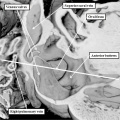
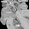
 Human cochlea stria vascularis 01.jpg 1,854 × 1,806; 754 KB
 Human fetal neural aneuploidy.jpg 1,000 × 1,400; 134 KB
 Human MII oocyte 01.jpg 1,200 × 826; 92 KB
 Human MII oocyte 02.jpg 1,200 × 826; 98 KB
 Human ovary follicle basement membrane 01.jpg 660 × 800; 184 KB
 Human spermatozoa acrosomal protein SP-10.jpg 1,100 × 1,189; 239 KB
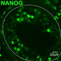
 Human testis NANOG expression.jpg 1,000 × 328; 77 KB
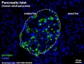 Human- pancreatic adult islet-glucagon.jpg 600 × 460; 67 KB
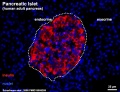 Human- pancreatic adult islet-insulin.jpg 600 × 460; 68 KB
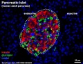 Human- pancreatic adult islet.jpg 600 × 460; 74 KB
 Human- spermatozoa NANOG expression 01.jpg 798 × 797; 79 KB
 Human- spermatozoa NANOG expression.jpg 1,000 × 333; 77 KB
 Meiotic prophase I stages.jpg 1,000 × 341; 66 KB
 Mitosis - Metaphase.jpg 384 × 384; 12 KB
 Mitosis fl.jpg 300 × 264; 21 KB
 Mitosis peroxisomes 01.jpg 1,200 × 394; 112 KB
 Mitosis peroxisomes 03.jpg 622 × 610; 61 KB
 Mitosis peroxisomes 04.jpg 622 × 610; 74 KB
 Mouse - forebrain Robo3 expression.jpg 675 × 1,280; 289 KB
 Mouse - palate MMP-25 expression.jpg 1,000 × 818; 243 KB
 Mouse blastocyst trophoblast 01.jpg 500 × 475; 22 KB
 Mouse gonad sex determination 01.jpg 600 × 600; 81 KB
 Mouse meiosis pachytene 01.jpg 1,280 × 317; 50 KB
- Mouse newborn ovary day 1.mp4 ; 646 KB
- Mouse newborn ovary day 2-3.5.mp4 ; 1.95 MB
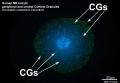
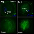
 Mouse oocyte cortical granules 01.jpg 500 × 475; 53 KB
 Mouse oocyte cortical granules 02.jpg 1,006 × 1,000; 177 KB
 Mouse oocyte microtubule-associated protein 01.jpg 869 × 1,000; 81 KB
 Mouse pancreas development.jpg 600 × 939; 261 KB
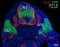 Mouse pax7 trunk neural crest 01.jpg 881 × 691; 158 KB
 Mouse pax7 trunk neural crest 02.jpg 881 × 691; 171 KB
 Mouse Wnt signaling 01.jpg 600 × 450; 156 KB
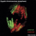 Mouse zygote mitosis anaphase.jpg 400 × 403; 24 KB
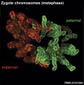 Mouse zygote mitosis metaphase.jpg 400 × 403; 30 KB
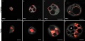
 Mouse zygote paternal genome reprogramming 01.jpg 1,157 × 1,280; 178 KB
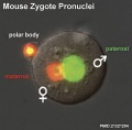 Mouse zygote pronuclei 01.jpg 800 × 784; 52 KB
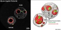 Mouse zygote pronuclei 02.jpg 1,000 × 501; 84 KB
 Mouse zygote pronuclei 03.jpg 1,200 × 586; 66 KB
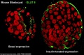 Mouse- blastocyst GLUT8 expression.jpg 789 × 519; 94 KB
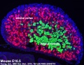 Mouse-adrenal gland E16.5.jpg 600 × 463; 93 KB
 Renal - early glomerulus.jpg 1,155 × 432; 52 KB
 Renal - podocyte development 01.jpg 1,200 × 908; 155 KB
 Renal - S-shaped body stage.jpg 1,155 × 432; 99 KB
 Transgenic rabbit.jpg 453 × 306; 28 KB
 Trisomy X.jpg 400 × 352; 15 KB
 Xenopus red fluorescence.jpg 600 × 873; 107 KB
 Zygote pronuclei stages 01.jpg 1,194 × 460; 49 KB


