Category:Dog
From Embryology
This page lists UNSW Embryology content related to dog development.
Pages in category 'Dog'
The following 17 pages are in this category, out of 17 total.
P
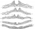
- Paper - Development and homology of the mammalian cerebellar fissures 2
- Paper - Development of the vascular system in five to twenty-one somite dog embryos
- Paper - Early differentiation of the foregut in the dog
- Paper - Malformation of the diaphragm in a dog (1924)
- Paper - On the prenatal and neonatal lung (1913)
Media in category 'Dog'
The following 53 files are in this category, out of 53 total.
 Alan Wilton.jpg 300 × 342; 40 KB
 Alaskan sled dogs.jpg 600 × 646; 97 KB
 Anson-1934 fig08-21.jpg 1,000 × 1,435; 178 KB
 Anson-1934 fig18.jpg 565 × 718; 36 KB
 Bailey064.jpg 790 × 597; 136 KB
 Bailey065.jpg 898 × 776; 145 KB
 Bailey070.jpg 717 × 1,112; 163 KB
 Bailey071.jpg 540 × 629; 59 KB
 Bailey120.jpg 891 × 599; 214 KB
 Bailey253.jpg 888 × 638; 123 KB
 Bailey254.jpg 734 × 808; 93 KB
 Bailey305.jpg 931 × 440; 82 KB
 Bailey370.jpg 975 × 1,084; 242 KB
 Bailey374.jpg 880 × 490; 75 KB
 Bailey488.jpg 942 × 433; 91 KB
 Canine embryo E21 image001.jpg 1,000 × 750; 152 KB
 Canine embryo E21 image002.jpg 600 × 450; 48 KB
 Canine embryo E35-38 image001.jpg 1,200 × 635; 199 KB
 Canine embryo E35-38 image002.jpg 1,000 × 774; 210 KB
 Canine embryo E35-38 image003.jpg 1,000 × 750; 192 KB
 Canine embryo E35-38 image004.jpg 613 × 1,000; 169 KB
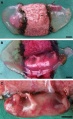
 Canine hepatozoonosis.jpg 600 × 490; 34 KB
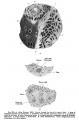
 Canine oocyte 01.jpg 624 × 532; 51 KB
 Canine oocyte 02.jpg 600 × 596; 43 KB
 Canine oocyte 03.jpg 600 × 694; 169 KB
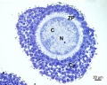 Canine oocyte 04.jpg 600 × 482; 76 KB
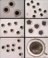 Canine oocyte to blastocyst.jpg 825 × 1,000; 154 KB
 Cloned dog.jpg 1,264 × 575; 89 KB
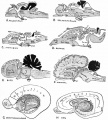
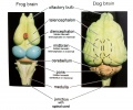 Comparative brain anatomy frog-dog.jpg 1,000 × 835; 112 KB
 Control and parthenogenetic canine fetuses.jpg 660 × 800; 111 KB
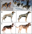
 Dog breeds 01.jpg 600 × 514; 35 KB
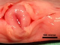
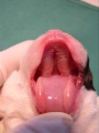 Dog day0-cleft palate.jpg 490 × 653; 39 KB
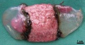
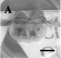 Dog fetus day 28.jpg 400 × 375; 24 KB
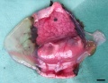
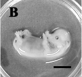 Dog fetus day 30.jpg 400 × 375; 32 KB
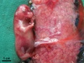
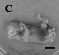 Dog fetus day 32.jpg 400 × 375; 28 KB
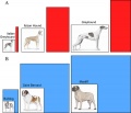
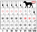
 Dog genetics.jpg 600 × 651; 108 KB
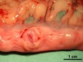
 Dog liver portosystemic shunts.jpg 800 × 264; 19 KB
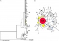 Dog mitochondrial DNA variations.jpg 1,000 × 703; 76 KB
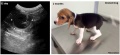
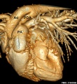 Dog patent ductus arteriosus computed tomography.jpg 600 × 654; 101 KB
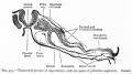
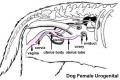 Dog- female urogenital cartoon.jpg 600 × 402; 28 KB
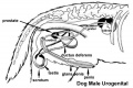 Dog- male urogenital cartoon.jpg 600 × 402; 30 KB
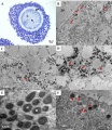
 Dog- spermatozoa NANOG expression.jpg 800 × 691; 109 KB
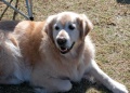 Dog-adult.jpg 500 × 360; 65 KB
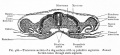
 Gray0015.jpg 800 × 682; 111 KB
 Keibel Mall 223.jpg 727 × 1,100; 89 KB
 Meyer1914 fig03.jpg 831 × 800; 69 KB
 Meyer1914 fig12.jpg 854 × 800; 269 KB
 Prentiss1906 fig02.jpg 1,224 × 891; 159 KB
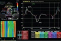 Speckle Tracking Echocardiograph of a dog affected with DMD.JPG 651 × 441; 67 KB
 Stricht-plate01.jpg 1,156 × 1,500; 243 KB
 Stricht-plate02.jpg 1,016 × 1,261; 308 KB
 Stricht-plate03.jpg 1,023 × 1,260; 230 KB
 Stricht-plate04.jpg 1,013 × 1,251; 238 KB


