Category:2011ANAT2341: Difference between revisions
From Embryology
No edit summary |
No edit summary |
||
| Line 1: | Line 1: | ||
UNSW Undergraduate Science course 2011 '''ANAT2341 Embryology''' content pages. | UNSW Undergraduate Science course 2011 '''ANAT2341 Embryology''' content pages. | ||
Revision as of 18:13, 28 July 2011
UNSW Undergraduate Science course 2011 ANAT2341 Embryology content pages.
Pages in category '2011ANAT2341'
The following 146 pages are in this category, out of 146 total.
2
- 2011 Group Project 1
- Talk:2011 Group Project 1
- 2011 Group Project 10
- Talk:2011 Group Project 10
- 2011 Group Project 11
- Talk:2011 Group Project 11
- 2011 Group Project 2
- Talk:2011 Group Project 2
- 2011 Group Project 3
- Talk:2011 Group Project 3
- 2011 Group Project 4
- Talk:2011 Group Project 4
- 2011 Group Project 5
- Talk:2011 Group Project 5
- 2011 Group Project 6
- Talk:2011 Group Project 6
- 2011 Group Project 7
- Talk:2011 Group Project 7
- 2011 Group Project 8
- Talk:2011 Group Project 8
- 2011 Group Project 9
- Talk:2011 Group Project 9
- 2011 Lab 1
- 2011 Lab 1 - Fertilization
- 2011 Lab 1 - Gametogenesis
- 2011 Lab 1 - Online Assessment
- 2011 Lab 1 - Oogenesis
- 2011 Lab 1 - Spermatogenesis
- 2011 Lab 10
- 2011 Lab 10 - Abnormalities
- 2011 Lab 10 - Early Embryo
- 2011 Lab 10 - Fetal
- 2011 Lab 10 - Late Embryo
- 2011 Lab 10 - Online Assessment
- 2011 Lab 10 - Postnatal
- 2011 Lab 11
- 2011 Lab 12
- Talk:2011 Lab 12
- 2011 Lab 12 - Abnormalities
- 2011 Lab 12 - Birth
- 2011 Lab 12 - Embryo to Fetus
- 2011 Lab 12 - Neonatal
- 2011 Lab 12 - Online Assessment
- 2011 Lab 12 - Second Trimester
- 2011 Lab 12 - Third Trimester
- 2011 Lab 2
- Talk:2011 Lab 2
- 2011 Lab 2 - Group Project
- 2011 Lab 2 - Online Assessment
- 2011 Lab 2 - Week 1
- 2011 Lab 2 - Week 2
- 2011 Lab 2 - Week 3
- 2011 Lab 3 - Group Project
- 2011 Lab 4
- 2011 Lab 5
- 2011 Lab 5 - Abnormalities
- 2011 Lab 5 - Early Embryo
- 2011 Lab 5 - Fetal
- 2011 Lab 5 - Gastrointestinal - Quiz
- 2011 Lab 5 - Late Embryo
- 2011 Lab 5 - Online Assessment
- 2011 Lab 5 - Postnatal
- 2011 Lab 5 - Trilaminar Embryo
- 2011 Lab 6
- 2011 Lab 6 - Abnormalities
- 2011 Lab 6 - Early Embryo
- 2011 Lab 6 - Fetal
- 2011 Lab 6 - Late Embryo
- 2011 Lab 6 - Online Assessment
- 2011 Lab 6 - Postnatal
- 2011 Lab 6 - Trilaminar Embryo
- 2011 Lab 8
- 2011 Lab 8 - Early Embryo
- 2011 Lab 8 - Fetal
- 2011 Lab 8 - Genital - Quiz
- 2011 Lab 8 - Genital Abnormalities
- 2011 Lab 8 - Late Embryo
- 2011 Lab 8 - Online Assessment
- 2011 Lab 8 - Postnatal
- 2011 Lab 8 - Sex Determination
- 2011 Lab 9
- Template:2011 Student Image
- Template:2011ANAT2341
- Template:2011Gp30Sep
- Template:2011GroupDiscussionMH
- Template talk:2011GroupDiscussionMH
- Template:2011Lab1
- Template:2011Lab10
- Template:2011Lab12
- Template:2011Lab2
- Template:2011Lab5
- Template:2011Lab5Footer
- Template:2011Lab6
- Template:2011Lab8
- Template:2011Projects
- Template:2011ProjectsMH
- Template:2011Student
A
Z
- User:Z3060621
- User:Z3217043
- User:Z3217345
- User:Z3272325
- User:Z3279511
- User:Z3284061
- User:Z3288196
- User:Z3288729
- User:Z3288827
- User:Z3289066
- User:Z3289301
- User:Z3289829
- User:Z3289991
- User:Z3290379
- User:Z3290558
- User:Z3290618
- User:Z3290689
- User:Z3290808
- User:Z3290815
- User:Z3291317
- User:Z3291324
- User:Z3291622
- User:Z3291643
- User:Z3292953
- User talk:Z3293267
- User:Z3308965
- User:Z3329495
- User:Z3332178
- User:Z3332183
- User:Z3332250
- User:Z3332327
- User:Z3387190
- User:Z3389343
- User:Z3389806
- User:Z3391078
Media in category '2011ANAT2341'
The following 200 files are in this category, out of 257 total.
(previous page) (next page) 2011 Project Group 1 edits.jpg 631 × 519; 28 KB
 2011 Project Group 1-11 edits.jpg 671 × 538; 40 KB
 2011 Project Group 10 edits.jpg 644 × 530; 28 KB
 2011 Project Group 11 edits.jpg 637 × 508; 31 KB
 2011 Project Group 2 edits.jpg 614 × 549; 30 KB
 2011 Project Group 3 edits.jpg 597 × 558; 31 KB
 2011 Project Group 4 edits.jpg 571 × 539; 29 KB
 2011 Project Group 5 edits.jpg 589 × 561; 30 KB
 2011 Project Group 6 edits.jpg 580 × 545; 30 KB
 2011 Project Group 7 edits.jpg 587 × 584; 34 KB
 2011 Project Group 8 edits.jpg 653 × 587; 33 KB
 2011 Project Group 9 edits.jpg 616 × 563; 33 KB
 2011 Talk Group 1 edits.jpg 600 × 410; 23 KB
 2011 Talk Group 1-11 edits.jpg 818 × 582; 44 KB
 2011 Talk Group 10 edits.jpg 595 × 431; 26 KB
 2011 Talk Group 11 edits.jpg 603 × 448; 28 KB
 2011 Talk Group 2 edits.jpg 601 × 400; 25 KB
 2011 Talk Group 3 edits.jpg 601 × 403; 25 KB
 2011 Talk Group 4 edits.jpg 598 × 402; 23 KB
 2011 Talk Group 5 edits.jpg 599 × 412; 24 KB
 2011 Talk Group 6 edits.jpg 599 × 393; 23 KB
 2011 Talk Group 7 edits.jpg 601 × 407; 25 KB
 2011 Talk Group 8 edits.jpg 599 × 432; 25 KB
 2011 Talk Group 9 edits.jpg 598 × 431; 25 KB
 22+23=45.jpg 594 × 158; 11 KB
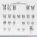 47,XXY Klinefelter's Syndrome.jpg 576 × 576; 58 KB
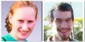
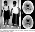
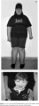 A 12 year old PWS patient and a 4 year old AS patient.jpg 358 × 966; 141 KB
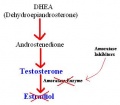 Action of Amoratase Inhibitors on Production of Estradiol.JPG 316 × 276; 12 KB
 Angelman Syndrome patient.png 343 × 271; 88 KB
 Angelo DiGeorge.png 331 × 480; 170 KB
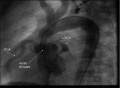 Angiography image indicating Supravalvular aortic stenosis.jpg 788 × 577; 133 KB
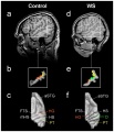 Auditory Cortex Location - Comparison Between Control Subject and WS Subject.jpg 1,084 × 1,261; 234 KB
 Bilateral Cleft Lip Variations.jpg 619 × 714; 87 KB
 Bilateral cleft lip with cleft hard and soft palate.jpg 168 × 137; 5 KB
 Bilateral cleft lip with cleft hard palate.jpg 167 × 141; 5 KB
 Bilateral Cleft Lip With Nasal Deformity.jpg 685 × 539; 148 KB
 Bilateral cleft lip.jpg 169 × 137; 5 KB
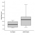 Blood Loss During Lip and Palate Repair.jpg 600 × 606; 21 KB
 Blood test result for glucose and iron.jpg 879 × 345; 54 KB
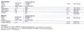 Blood test results.jpg 879 × 345; 54 KB
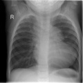 Chestxrayfallot.jpg 630 × 630; 136 KB
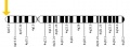
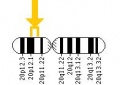
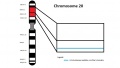 Chromosome 20 - JAG1 gene.jpg 976 × 552; 55 KB
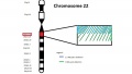 Chromosome 22 - TBX1 Gene.jpg 960 × 540; 71 KB
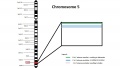 Chromosome 5 - NKX2-5 Gene.jpg 960 × 540; 70 KB
 Chromosome 7, indicating 7q11.23 region of Williams Syndrome.gif 325 × 270; 11 KB
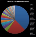
 Cleft Palate Maxillary and Mandibular View.jpg 547 × 658; 141 KB
 Clinical examination TOF.png 479 × 350; 347 KB
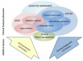
 Cognitive performance in WS subjects (n = 67) versus normal controls.png 600 × 292; 139 KB
 Comparison of morphogenesis of the upper lip with the palate.jpg 774 × 371; 188 KB
 Control group response to startle.jpg 661 × 492; 66 KB
 Critical region of Angelman Syndrome on chromosome 15.png 591 × 576; 356 KB
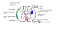 Cross Section of the Spinal Cord.jpg 1,280 × 640; 74 KB
 CT lungcancer.JPG 422 × 348; 18 KB
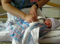
 Cyanotic baby.jpg 469 × 600; 43 KB
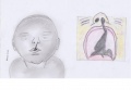
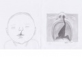
 Development 2.jpg 588 × 637; 22 KB
 Development 3.jpg 588 × 637; 21 KB
 Development1.jpg 588 × 637; 22 KB
 DiGeorge Baby.jpg 691 × 800; 64 KB
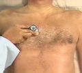
 Doctor performing an ECG on patient.png 539 × 334; 304 KB
 Dr Charles Williams.jpg 430 × 500; 101 KB
 Dr Etienne Louis Arthur Fallot.jpg 434 × 576; 53 KB
 Dr Harry Angelman.jpg 481 × 600; 49 KB
 Dr Helen Brooke Taussig.jpg 307 × 418; 29 KB
 Duchenne.JPG 324 × 457; 22 KB
 Dystrophin in the muscle fibre membrane.jpg 1,840 × 1,355; 415 KB
 Dystrophin within the plasma membrane of muscle fibres.jpg 800 × 640; 112 KB
 Echocardiogram concentric left ventricular hypertrophy.jpg 800 × 460; 55 KB
 Effect of Frataxin Levels.jpg 1,360 × 624; 84 KB
 Electrocardiograph findings in dogs affected with DMD.JPG 609 × 538; 64 KB
 Electroencephalography of Angelman Syndrome.jpg 440 × 207; 33 KB
 Embryo2011-Ectoderm 110811 part 1.mov ; 6.04 MB
 Embryo2011-Ectoderm 110811 part 1.mp3 ; 6.03 MB
- Embryo2011-Ectoderm 110811 part 2.mov ; 6.56 MB
- Embryo2011-Ectoderm 110811 part 2.mp3 ; 6.55 MB
- Embryo2011-GIT 230811 part 1.mov ; 6.44 MB
- Embryo2011-GIT 230811 part 2.mov ; 6.45 MB
- Embryo2011-Head 300811 part 1.mov ; 6.5 MB
- Embryo2011-Head 300811 part 1.mp3 ; 6.77 MB
- Embryo2011-Head 300811 part 2.mov ; 6.4 MB
- Embryo2011-Head 300811 part 2.mp3 ; 6.66 MB
- Embryo2011-Mesoderm 090811 part 1.mov ; 6.12 MB
- Embryo2011-Mesoderm 090811 part 2.mov ; 5.86 MB
- Embryo2011-Mesoderm 090811 part 2.mp3 ; 5.85 MB
- Embryo2011-Placenta 180811 part 1.mov ; 6.37 MB
- Embryo2011-Placenta 180811 part 1.mp3 ; 6.36 MB
- Embryo2011-Placenta 180811 part 2.mov ; 6.25 MB
- Embryo2011-Placenta 180811 part 2.mp3 ; 6.23 MB
- Embryo2011-Week 3 040811 part 1.mov ; 6.05 MB
- Embryo2011-Week 3 040811 part 1.mp3 ; 6.04 MB
- Embryo2011-Week 3 040811 part 2.mov ; 6.72 MB
- Embryo2011-Week 3 040811 part 2.mp3 ; 6.7 MB
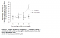 Emotional Response to 'cartoon' test Klinefelter Syndrome.png 1,008 × 630; 174 KB
 Extent of microcephaly in Angelman Syndrome patients.png 1,000 × 814; 43 KB
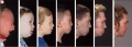
 Facial features of four individuals with Willams Syndrome.gif 429 × 182; 45 KB
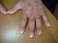 Finger-Clubbing.jpg 600 × 449; 58 KB
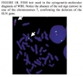 FISH test used to confirm the deletion of the ELN gene.jpg 337 × 312; 15 KB
 Fluorescence In situ Hybridization (FISH) assay.JPG 696 × 222; 11 KB
 FMR1 is silenced in FXS.jpg 451 × 497; 21 KB
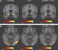 FMRI Images of Brain Activation in XXY Patients.JPG 433 × 378; 23 KB
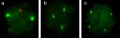
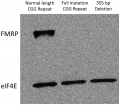 FMRP expression in control and fragile X tissues.png 600 × 527; 1.29 MB
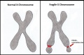 Fragile X Chromosome..jpg 997 × 656; 69 KB
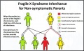
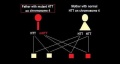
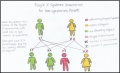 Fragile x inheritance..jpg 1,074 × 654; 66 KB
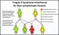 Fragile X Inheritance..jpg 1,074 × 654; 105 KB
 Fragile x inheritance.jpg 1,074 × 654; 102 KB
 Fragile X Phenotype.jpg 225 × 112; 23 KB
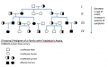
 Friedreich's Ataxia Pedigree.jpg 1,424 × 588; 80 KB
 Friedreich's Ataxia Pedigree.png 618 × 384; 58 KB
 From infancy until completion of treatment.jpg 702 × 254; 103 KB
 Furlow Z-plasty technique.jpg 670 × 771; 147 KB
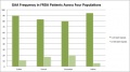 GAA Frequency in FRDA.jpg 622 × 345; 37 KB
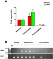
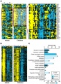 Gene expression responses of Friedreich's ataxia.jpg 551 × 749; 342 KB
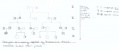
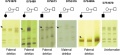 Genotyping of the five microsatellites markers in WBS families.jpg 465 × 213; 71 KB
 George Huntington.jpg 285 × 358; 44 KB
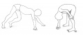 Gower's sign - a symptom of DMD.JPG 1,144 × 491; 36 KB
 HD future research.jpg 687 × 869; 78 KB
 HD Interview questions.png 204 × 135; 12 KB
- HD patient with no treatment.mov ; 1.17 MB
 HD patients.jpg 492 × 442; 52 KB
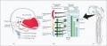
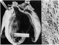 Heart disection.jpg 963 × 731; 209 KB
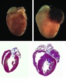 Heart Hypertrophy gross.jpg 639 × 800; 59 KB
 Heart murmur TOF.png 385 × 343; 172 KB
 Hippocampal formation.pdf ; 149 KB
 House drawings Williams.jpg 2,328 × 1,464; 112 KB
 Huntingtin gene.jpeg 492 × 181; 15 KB
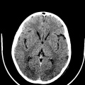 Huntington disease atrophy 1.jpg 630 × 630; 148 KB
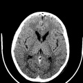 Huntington disease atrophy 2.jpg 630 × 630; 146 KB
 Huntington disease atrophy 3.jpg 630 × 630; 144 KB
 Huntington Disease patient and control MRI.gif 530 × 352; 139 KB
 Huntington's disease MRI.jpg 504 × 630; 163 KB
 Imprint defect inheritance in Angelman Syndrome.png 688 × 323; 21 KB
 Inheritance pattern in Huntington's Disease.jpeg 531 × 284; 15 KB
 Introduction 3.jpg 921 × 637; 40 KB
 Introduction 4.jpg 907 × 637; 31 KB
 JAG1.jpeg 196 × 139; 6 KB
 Karyotype of a Klinefelter's syndrome patient.jpg 779 × 545; 85 KB
 Karyotype of Klinefelter's Syndrome.png 768 × 600; 222 KB
 Karyotype.jpg 648 × 454; 49 KB
 Key cellular pathogenic mechanisms in HD.jpg 663 × 517; 47 KB
 Key cellular pathogenic mechanisms in Huntington's disease.jpg 663 × 517; 47 KB
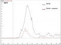 Klinefelter syndrome group response to startle.jpg 661 × 492; 73 KB
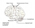 Lateral View of the Brain.jpeg 782 × 627; 102 KB
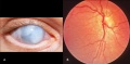 LCA fundus and cataracts.jpg 570 × 280; 149 KB
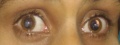 LCA patient.jpg 464 × 175; 78 KB
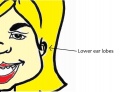 Lowered Ear Lobes.jpg 1,078 × 828; 72 KB
 Magnetic Resonance Imaging in an adult patient with tof.JPG 188 × 189; 7 KB
 Major causes of death in surgically untreated TOF patients.png 1,009 × 570; 127 KB
 Maternal Non-Disjunction.PNG 952 × 525; 52 KB
 Mechanism of tetrabenazine.jpg 600 × 459; 33 KB
 Median facial dysplasia.jpg 597 × 448; 179 KB
 Meiotic non-disjunction.jpg 989 × 299; 35 KB
 Mice mutants exhibit cleft palate and umbilical hernia.jpg 527 × 332; 126 KB
 Migration.jpg 771 × 333; 118 KB
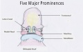 Modified prominences final.jpg 667 × 423; 23 KB
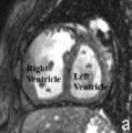
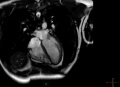 MRI heart.JPG 491 × 354; 14 KB
 MSX 1 Gene.JPG 385 × 677; 22 KB
 Neuroacanthocytosis.jpg 487 × 584; 91 KB
 NeuromericOrganization.jpg 620 × 449; 123 KB
 Nikolaus Friedreich Portrait.jpg 320 × 576; 54 KB
 NKX2-5.jpeg 469 × 173; 14 KB
 Nondisjunction of Homologous Chromosomes in Meiosis1.jpg 900 × 636; 113 KB
 Nondisjunction of Sister Chromatids in Meiosis 2.jpg 900 × 636; 121 KB
 Nondisjunction.jpg 638 × 135; 23 KB
 Normal and Angelman Syndrome mice models.jpg 700 × 505; 135 KB
 Normal fetal blood flow and Tetralogy of Fallot.jpg 628 × 543; 200 KB
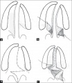
 Normal palate shelf and key stages of mouse palatal development.jpg 771 × 153; 80 KB
 Oral Clefting.JPG 662 × 464; 31 KB
 Pathogenesis of Friedreich Ataxia.jpg 520 × 380; 30 KB
 Pes Cavus Deformity.jpg 649 × 247; 79 KB
 Phenotypes of FXS overlap with those of Autism.jpg 600 × 441; 51 KB
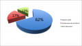
 Pie Chart.JPG 551 × 572; 50 KB
 Pierre Joseph Desault.png 1,431 × 2,100; 2.34 MB
 Point mutations resulting in DMD.jpg 2,051 × 1,131; 412 KB
 Point vs frameshift mutation of DMD gene.png 1,689 × 1,000; 874 KB
 Potts Shunt.jpg 311 × 456; 117 KB
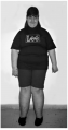 Prader-Willi Syndrome patient.png 343 × 650; 149 KB
 Psoriasis.jpg 275 × 181; 9 KB
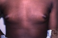 Pubertal gynecomastia 1.jpg 550 × 364; 92 KB


_versus_normal_controls.png)


_vs._Duchennes_muscular_dystrophy_muscle_(b).jpg)


_assay.JPG)