BGDA Practical Placenta - Diagnostic Techniques: Difference between revisions
No edit summary |
|||
| Line 30: | Line 30: | ||
{{Template:Prenatal diagnosis}} | [[Placenta_-_Membranes#Amnionic_Sac|Placenta - Amnionic Sac]] | {{Template:Prenatal diagnosis}} | [[Placenta_-_Membranes#Amnionic_Sac|Placenta - Amnionic Sac]] | ||
==Placenta Term Parameters== | |||
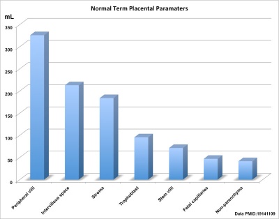
[[File:Placental_volume_graph.jpg|thumb|400px|Human term placental volumes sorted by size (from table).]] | |||
Term placental composition, villous capillarization and the mean cross-sectional areas of peripheral villi and capillaries, data from a study sample of 15 normal placenta (mean placental volume, 652 ml). <ref><pubmed>19141109</pubmed></ref> | |||
{| | |||
|-bgcolor="CEDFF2" | |||
| width="150px"|'''Variable''' | |||
| '''Unit''' | |||
| '''Placenta''' (mean, n = 15) | |||
|- | |||
|Intervillous space | |||
| mL | |||
| 213 | |||
|-bgcolor="F5FAFF" | |||
| Stem villi | |||
| mL | |||
| 71.4 | |||
|- | |||
| Peripheral villi | |||
| mL | |||
| 326 | |||
|-bgcolor="F5FAFF" | |||
| Trophoblast | |||
| mL | |||
| 95.5 | |||
|- | |||
| Stroma | |||
| mL | |||
| 184 | |||
|-bgcolor="F5FAFF" | |||
| Fetal capillaries | |||
| mL | |||
| 46.9 | |||
|- | |||
| Non-parenchyma | |||
| mL | |||
| 41.5 | |||
|-bgcolor="F5FAFF" | |||
| Peripheral villi | |||
| km | |||
| 89.2 | |||
|- | |||
| Fetal capillaries | |||
| km | |||
| 310 | |||
|-bgcolor="F5FAFF" | |||
| TS area villi | |||
| µm<sup>2</sup> | |||
| 3700 | |||
|- | |||
| TS area capillary | |||
| µm<sup>2</sup> | |||
| 150 | |||
|-bgcolor="F5FAFF" | |||
| Capillaries | |||
| mL mL<sup>-1</sup> | |||
| 0.147 | |||
|- | |||
| Length ratio | |||
| km km<sup>-1</sup> | |||
| 3.6 | |||
|-bgcolor="F5FAFF" | |||
|} | |||
<references/> | |||
==Terms== | ==Terms== | ||
Revision as of 19:26, 29 May 2012
| Practical 14: Implantation and Early Placentation | Villi Development | Maternal Decidua | Cord Development | Placental Functions | Diagnostic Techniques | Abnormalities |
Chorionic Villus Sampling

|
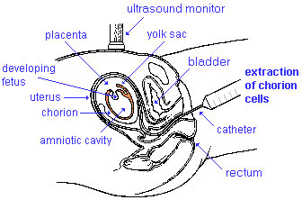
Chorionic Villus Sampling test is done in the 10th to 12th week after the first day of the mother's last menstrual period. The test is done by looking at cells taken from the chorionic membrane or placenta. No anaesthetic is required, and a test result is usually available in two to three weeks.
When the test is carried out by an obstetrician experienced in the technique, the risk of miscarriage related to the test is about 2%. Potential disadvantages include maternal cell contamination, placental mosaicism and failure to obtain an adequate specimen. This may result in the need for a repeat procedure or amniocentesis.
|
Amniocentesis

|
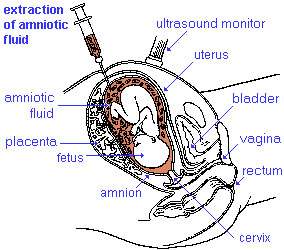
Amniocentesis is a prenatal diagnostic test carried out mainly between 14th to 18th week of pregnancy. Amniotic fluid is taken from the uterus, sent to a diagnostic laboratory and embryonic cells isolated from the amniotic fluid. No anaesthetic is required, and a result is usually obtained in about three to four weeks. When the test is carried out by an obstetrician experienced in the technique, the risk of a miscarriage related to the test is about 1 %.
|
Placenta Term Parameters
Term placental composition, villous capillarization and the mean cross-sectional areas of peripheral villi and capillaries, data from a study sample of 15 normal placenta (mean placental volume, 652 ml). [1]
| Variable | Unit | Placenta (mean, n = 15) |
| Intervillous space | mL | 213 |
| Stem villi | mL | 71.4 |
| Peripheral villi | mL | 326 |
| Trophoblast | mL | 95.5 |
| Stroma | mL | 184 |
| Fetal capillaries | mL | 46.9 |
| Non-parenchyma | mL | 41.5 |
| Peripheral villi | km | 89.2 |
| Fetal capillaries | km | 310 |
| TS area villi | µm2 | 3700 |
| TS area capillary | µm2 | 150 |
| Capillaries | mL mL-1 | 0.147 |
| Length ratio | km km-1 | 3.6 |
- ↑ <pubmed>19141109</pubmed>
Terms
BGDA: Lecture 1 | Lecture 2 | Practical 3 | Practical 6 | Practical 12 | Lecture Neural | Practical 14 | Histology Support - Female | Male | Tutorial
Glossary Links
- Glossary: A | B | C | D | E | F | G | H | I | J | K | L | M | N | O | P | Q | R | S | T | U | V | W | X | Y | Z | Numbers | Symbols | Term Link
Cite this page: Hill, M.A. (2024, May 13) Embryology BGDA Practical Placenta - Diagnostic Techniques. Retrieved from https://embryology.med.unsw.edu.au/embryology/index.php/BGDA_Practical_Placenta_-_Diagnostic_Techniques
- © Dr Mark Hill 2024, UNSW Embryology ISBN: 978 0 7334 2609 4 - UNSW CRICOS Provider Code No. 00098G

