ANAT2341 Lab 4 - Implantation and Villi Development
| ANAT2341 Lab 4: Introduction | Implantation and Villi | Decidua and Cord | Abnormal Placenta | Cardiovascular | Online Assessment | Group Project |
Implantation

Human Embryo Day 8 to 9
| Implantation | Chorionic Cavity | Week 3 | Amniotic Cavity |
| Quicktime | Flash | Quicktime | Flash | Quicktime | Flash | Quicktime | Flash |
Week 3 embryo


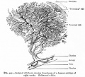
Villi Development
Chorionic Villi
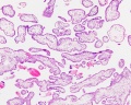
| Primary villi
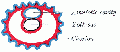
Week 2 - first stage of chorionic villi development, trophoblastic shell cells (syncitiotrophoblasts and cytotrophoblasts) form finger-like extensions into maternal decidua. |

|
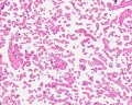
| Secondary villi
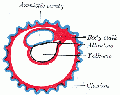
Week 3 - second stage of chorionic villi development, extraembryonic mesoderm grows into villi, covers entire surface of chorionic sac. Basal region will form chorionic plate. |

|
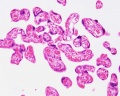
| Tertiary villi
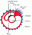
Week 4 - third stage of chorionic villi development, mesenchyme differentiates into blood vessels and cells, forms arteriocapillary network, fuse with placental vessels, developing in connecting stalk.
|

|
Anchoring Villi

Floating Villi


Placenta anchoring villi
Villi first trimester
Villi at term
Villi at term


Chorionoic Villi Location
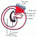
Originally villi cover entire chorionic surface and then become restricted to decidua basalis region forming 2 regions:
- Frondosum - "leafy" where villi are mainly located.
- Capsularis - smooth chorion, where villi are absent or not abundant.

|

|
| ANAT2341 Lab 4: Introduction | Implantation and Villi | Decidua and Cord | Abnormal Placenta | Cardiovascular | Online Assessment | Group Project |
Additional Information
Human Villi Timeline
The placental vill development data below is based upon a recent immunochemistry confocal laser scanning microscope (CLSM) study.[1]
Note that the paper uses clinical gestational age (GA) from last menstrual period (LMP) and has been corrected for post-conception (fertilization) age, approximately 14 days later.
| Fertilization Age
(weeks) |
Gestational Age
(weeks) |
Vessel Lumen Diameter
(range in microns, µm) |
Features |
| 3 to 4 | 5 and 6 | 10 - 15 |
|
| 5 to 6 | 7 and 8 | 10 - 26 |
|
| 7 to 8 | 9 and 10 | 60 - 75 two central vessels
26 - 34 capillary network |
|
| 9 to 10 | 11 and 12 | 70 - 90 two central vessels
26 - 34 capillary network |
|
Reference
- ↑ <pubmed>17545656</pubmed>
Decidua and villi location
Chorionic villi


| ANAT2341 Lab 4: Introduction | Implantation and Villi | Decidua and Cord | Abnormal Placenta | Cardiovascular | Online Assessment | Group Project |
- 2012 Course: Week 1 Lecture 1 Lecture 2 Lab 1 | Week 2 Lecture 3 Lecture 4 Lab 2 | Week 3 Lecture 5 Lecture 6 Lab 3 | Week 4 Lecture 7 Lecture 8 Lab 4 | Week 5 Lecture 9 Lecture 10 Lab 5 | Week 6 Lecture 11 Lecture 12 Lab 6 | Week 7 Lecture 13 Lecture 14 | Lab 7 | Week 8 Lecture 15 Lecture 16 Lab 8 | Week 9 Lecture 17 Lecture 18 Lab 9 | Week 10 Lecture 19 Lecture 20 Lab 10 | Week 11 Lecture 21 Lecture 22 Lab 11 | Week 12 Lecture 23 Lecture 24 Lab 12
Glossary Links
- Glossary: A | B | C | D | E | F | G | H | I | J | K | L | M | N | O | P | Q | R | S | T | U | V | W | X | Y | Z | Numbers | Symbols | Term Link
Cite this page: Hill, M.A. (2024, June 15) Embryology ANAT2341 Lab 4 - Implantation and Villi Development. Retrieved from https://embryology.med.unsw.edu.au/embryology/index.php/ANAT2341_Lab_4_-_Implantation_and_Villi_Development
- © Dr Mark Hill 2024, UNSW Embryology ISBN: 978 0 7334 2609 4 - UNSW CRICOS Provider Code No. 00098G