File:Infant lymphocytic choriomeningitis virus CT.jpg
Original file (665 × 800 pixels, file size: 69 KB, MIME type: image/jpeg)
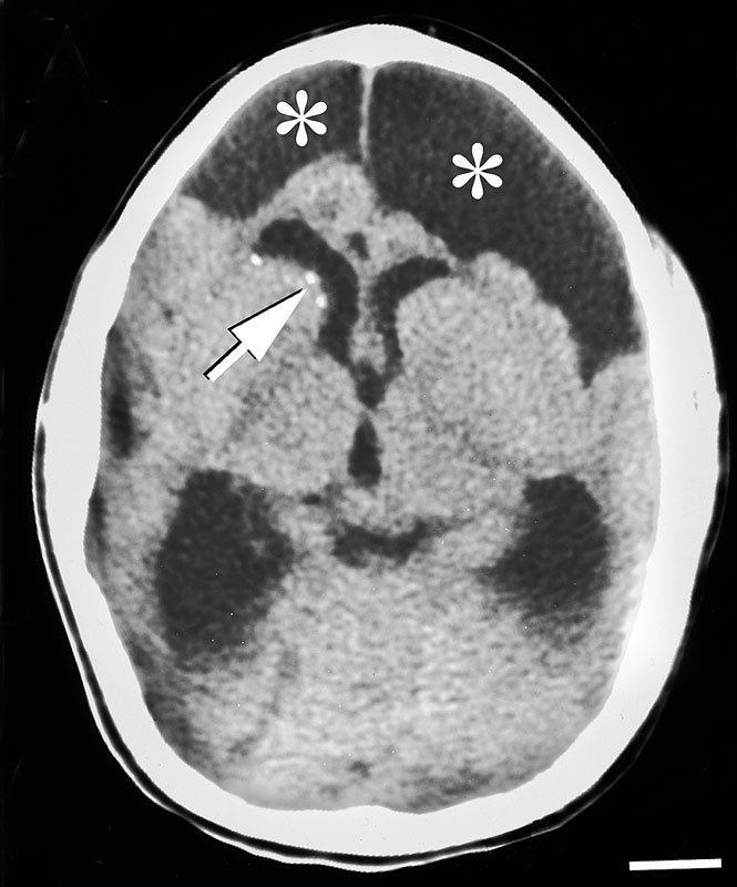
Congenital Lymphocytic choriomeningitis virus LCMV infection Computed Tomography
Head CT scan from a 4 month old child with congenital LCMV infection.
The scan reveals bilateral asymmetric regions of encephalomalacia (asterisks), strongly suggestive of a focal destructive process. Note also the periventricular calcifications (arrow), characterisitic of a prenatal viral infection.
X-ray computed tomography, also Computed tomography (CT scan)
Reference
PMID 18052527
http://www.plospathogens.org/article/info%3Adoi%2F10.1371%2Fjournal.ppat.0030149
PLoS Pathog. 2007 Nov;3(11):e149. Congenital viral infections of the brain: lessons learned from lymphocytic choriomeningitis virus in the neonatal rat. Bonthius DJ, Perlman S. Source Department of Pediatrics, Neurology, and Anatomy at the Carver College of Medicine, University of Iowa, Iowa City, Iowa, United States of America. daniel-bonthius@uiowa.edu Abstract The fetal brain is highly vulnerable to teratogens, including many infectious agents. As a consequence of prenatal infection, many children suffer severe and permanent brain injury and dysfunction. Because most animal models of congenital brain infection do not strongly mirror human disease, the models are highly limited in their abilities to shed light on the pathogenesis of these diseases. The animal model for congenital lymphocytic choriomeningitis virus (LCMV) infection, however, does not suffer from this limitation. LCMV is a well-known human pathogen. When the infection occurs during pregnancy, the virus can infect the fetus, and the developing brain is particularly vulnerable. Children with congenital LCMV infection often have substantial neurological deficits. The neonatal rat inoculated with LCMV is a superb model system of human congenital LCMV infection. Virtually all of the neuropathologic changes observed in humans congenitally infected with LCMV, including microencephaly, encephalomalacia, chorioretinitis, porencephalic cysts, neuronal migration disturbances, periventricular infection, and cerebellar hypoplasia, are reproduced in the rat model. Within the developing rat brain, LCMV selectively targets mitotically active neuronal precursors. Thus, the targets of infection and sites of pathology depend on host age at the time of infection. The rat model has further shown that the pathogenic changes induced by LCMV infection are both virus-mediated and immune-mediated. Furthermore, different brain regions simultaneously infected with LCMV can undergo widely different pathologic changes, reflecting different brain region-virus-immune system interactions. Because the neonatal rat inoculated with LCMV so faithfully reproduces the diverse neuropathology observed in the human counterpart, the rat model system is a highly valuable tool for the study of congenital LCMV infection and of all prenatal brain infections In addition, because LCMV induces delayed-onset neuronal loss after the virus has been cleared, the neonatal rat infected with LCMV may be an excellent model system to study neurodegenerative or psychiatric diseases whose etiologies are hypothesized to be virus-induced, such as autism, schizophrenia, and temporal lobe epilepsy.
PMID 18052527
Copyright: © 2007 Bonthius and Perlman. This is an open-access article distributed under the terms of the Creative Commons Attribution License, which permits unrestricted use, distribution, and reproduction in any medium, provided the original author and source are credited.
Journal.ppat.0030149.g001.jpg
File history
Click on a date/time to view the file as it appeared at that time.
| Date/Time | Thumbnail | Dimensions | User | Comment | |
|---|---|---|---|---|---|
| current | 04:58, 1 June 2012 | | 665 × 800 (69 KB) | Z8600021 (talk | contribs) | ==Congenital Lymphocytic choriomeningitis virus LCMV infection Computed Tomography== Head CT scan from a 4 month old child with congenital LCMV infection. The scan reveals bilateral asymmetric regions of encephalomalacia (asterisks), strongly suggestiv |
You cannot overwrite this file.
File usage
The following page uses this file: