File:Nephrons-cortical and juxtamedullary.jpg
Nephrons-cortical_and_juxtamedullary.jpg (507 × 600 pixels, file size: 39 KB, MIME type: image/jpeg)
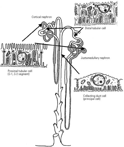
Schematic of two nephrons (cortical and juxtamedullary). The cell types shown are restricted to those most widely used for either isolation or establishment of primary cultures: a proximal tubular cell from the convoluted portion, a cell from the distal convoluted tubule, and a principal cell from the cortical collecting duct.
Original Image name: Pfallerfig1-600px.jpg
Image Source: Alternative Testing Methodologies - Nephrotoxicity Testing in Vitro--What We Know and What We Need to Know Environmental Health Perspectives, Volume 106, Supplement 2, April 1998 http://www.ehponline.org/realfiles/members/1998/Suppl-2/559-569pfaller/pfaller.html
File history
Click on a date/time to view the file as it appeared at that time.
| Date/Time | Thumbnail | Dimensions | User | Comment | |
|---|---|---|---|---|---|
| current | 15:17, 21 September 2009 | | 507 × 600 (39 KB) | S8600021 (talk | contribs) | Schematic of two nephrons (cortical and juxtamedullary). The cell types shown are restricted to those most widely used for either isolation or establishment of primary cultures: a proximal tubular cell from the convoluted portion, a cell from the distal c |
You cannot overwrite this file.
File usage
The following 3 pages use this file: