File:Early zygote labelled.jpg
Early_zygote_labelled.jpg (500 × 441 pixels, file size: 29 KB, MIME type: image/jpeg)
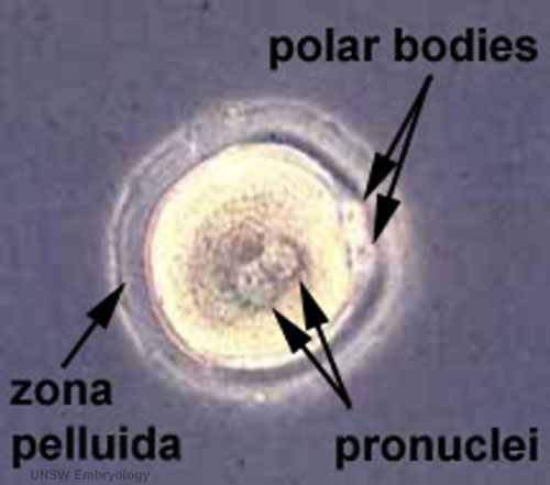
Early Zygote
This is described as an early human zygote due to the presence of pronuclei in the centre of the cytoplasm.
The haploid pronuclei of the male (spermatozoa) and female (oocyte) have not yet combined to form a single nucleus.
The polar bodies can be seen at the edge of the cytoplasm (at 3 o'clock position). These exclusion bodies contain the additional oocyte DNA produced in meiosis.
The zona pellucida forms the thick clear layer that surrounds the cell.
At this stage in vivo:
- there would still be granulosa cells and spermatozoa attached to the zone pellucida.
- the zygote floats freely within the uterine tube.
- The cell is preparing for the first mitotic division.
About Carnegie Stages 1
Facts: Week 1, size 0.1 - 0.15 mm (100 - 150 microns)
Features: zygote, fertilized oocyte, pronuclei, polar bodies, zona pellucida
Image Source: UNSW Embryology http://embryology.med.unsw.edu.au/wwwhuman/Stages/Stage1.htm
File history
Click on a date/time to view the file as it appeared at that time.
| Date/Time | Thumbnail | Dimensions | User | Comment | |
|---|---|---|---|---|---|
| current | 11:47, 10 March 2012 | | 500 × 441 (29 KB) | Z8600021 (talk | contribs) | increase image size |
| 13:28, 20 July 2010 | 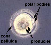 | 216 × 191 (8 KB) | S8600021 (talk | contribs) | ==Early Zygote (labelled)== About Carnegie Stages 1 Facts: Week 1, size 0.1-0.15 mm Features: zygote, fertilized oocyte, pronuclei, polar bodies, zona pellucida Related Images: Early zygote | Image Source: UNSW Embryology ht |
You cannot overwrite this file.
File usage
The following 14 pages use this file:
- 2015 Group Project 1
- BGDA Lecture - Development of the Embryo/Fetus 1
- Carnegie stage 1
- Embryonic Development
- Fertilization
- Lecture - 2015 Course Introduction
- Lecture - 2016 Course Introduction
- Lecture - 2017 Course Introduction
- Lecture - Fertilization
- P
- Z
- Zygote
- Talk:2015 Group Project 1
- Talk:Lecture - 2016 Course Introduction