File:Skull CT normal sutures 01.jpg
Original file (1,000 × 526 pixels, file size: 89 KB, MIME type: image/jpeg)
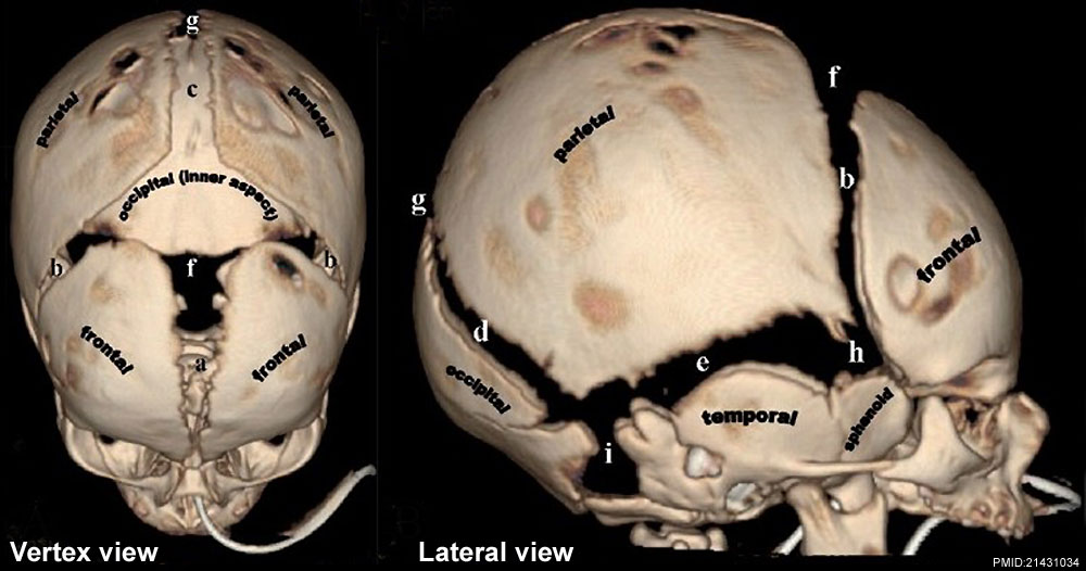
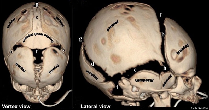
Skull Normal Sutures
Computed Tomography (CT) scan with 3D surface-rendered reconstructions.
A - Vertex view
B - Lateral view
- (a) Metopic suture; (b) coronal sutures; (c) sagittal suture; (d) lambdoid suture; (e) squamosal suture; (f) anterior fontanel; (g) posterior fontanel; (h) sphenoidal fontanel; (i) mastoid fontanel.
- Cranial vault bones usually ossify from the center to periphery, which results in this “widened” appearance of the sutures in the newborn.
- Links: Skull Development | Historic - skull of a human fetus of 43 millimeters greatest length | Computed Tomography
Reference
<pubmed>21431034</pubmed>| PMC3056371 | Indian J Radiol Imaging.
This is an open-access article distributed under the terms of the Creative Commons Attribution License, which permits unrestricted use, distribution, and reproduction in any medium, provided the original work is properly cited.
Paritosh C Khanna © 2007 - 2012 Indian Journal of Radiology and Imaging
Attribution-NonCommercial-ShareAlike 3.0 Unported (CC BY-NC-SA 3.0)
Original file name: Figure 1(A-E): IJRI-21-49-g001.jpg
http://www.ijri.org/viewimage.asp?img=IndianJRadiolImaging_2011_21_1_49_76055_f2.jpg
File history
Click on a date/time to view the file as it appeared at that time.
| Date/Time | Thumbnail | Dimensions | User | Comment | |
|---|---|---|---|---|---|
| current | 08:23, 17 March 2012 | | 1,000 × 526 (89 KB) | Z8600021 (talk | contribs) | ==Skull Normal Sutures== Computed Tomography (CT) scan with 3D surface-rendered reconstructions. ===A - Vertex view=== ===B - Lateral view=== * (a) Metopic suture; (b) coronal sutures; (c) sagittal suture; (d) lambdoid suture; (e) squamosal suture; (f |
You cannot overwrite this file.
File usage
The following 5 pages use this file: