File:Skull CT normal sutures 01.jpg
Original file (1,000 × 526 pixels, file size: 89 KB, MIME type: image/jpeg)
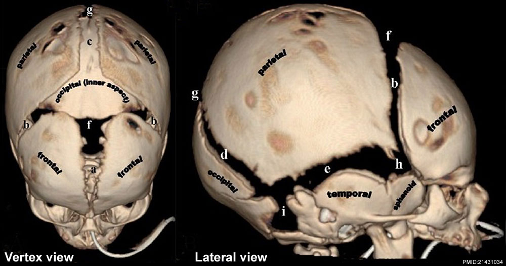
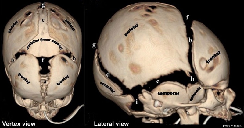
Skull Normal Sutures
Computed Tomography (CT) scan with 3D surface-rendered reconstructions.
A - Vertex view
B - Lateral view
- (a) Metopic suture; (b) coronal sutures; (c) sagittal suture; (d) lambdoid suture; (e) squamosal suture; (f) anterior fontanel; (g) posterior fontanel; (h) sphenoidal fontanel; (i) mastoid fontanel.
- Cranial vault bones usually ossify from the center to periphery, which results in this “widened” appearance of the sutures in the newborn.
C - Endocranial skull base view
Shows portions of the occipital bone and sutures
- (j) Basioccipital; (k) paired exoccipital; (l) supraoccipital; and (m) interparietal. Associated synchondroses are (n) spheno-occipital; (o)anterior intra-occipital; (p) posterior intra-occipital; (q) petro-occipital; (r) occipitomastoid; (s) and mendosal sutures. Note that o, k, p and s are paired structures.
D - Vertex view
Shows the lambda (point of intersection of the sagittal and lambdoid sutures) and bregma (point of intersection of the coronal and sagittal sutures.
E - Endocranial skull base view
Shows the basion (located on the basiocciput, at the midpoint of the anterior margin of the foramen magnum) and opisthion (located on the occipital bone, at the midpoint of the posterior margin of the foramen magnum).
- Links: Skull Development | Historic - skull of a human fetus of 43 millimeters greatest length | Computed Tomography
Reference
<pubmed>21431034</pubmed>| PMC3056371 | Indian J Radiol Imaging.
This is an open-access article distributed under the terms of the Creative Commons Attribution License, which permits unrestricted use, distribution, and reproduction in any medium, provided the original work is properly cited.
Paritosh C Khanna © 2007 - 2012 Indian Journal of Radiology and Imaging
Attribution-NonCommercial-ShareAlike 3.0 Unported (CC BY-NC-SA 3.0)
Original file name: Figure 1(A-E): IJRI-21-49-g001.jpg
http://www.ijri.org/viewimage.asp?img=IndianJRadiolImaging_2011_21_1_49_76055_f2.jpg
File history
Click on a date/time to view the file as it appeared at that time.
| Date/Time | Thumbnail | Dimensions | User | Comment | |
|---|---|---|---|---|---|
| current | 08:23, 17 March 2012 | | 1,000 × 526 (89 KB) | Z8600021 (talk | contribs) | ==Skull Normal Sutures== Computed Tomography (CT) scan with 3D surface-rendered reconstructions. ===A - Vertex view=== ===B - Lateral view=== * (a) Metopic suture; (b) coronal sutures; (c) sagittal suture; (d) lambdoid suture; (e) squamosal suture; (f |
You cannot overwrite this file.
File usage
The following 5 pages use this file: