Foundations - Histology Cells and Tissues
Introduction
Background and Self-directed Learning for Medicine Foundations.
Practical - Histology Cells and Tissues Virtual Slides by Patrick de Permentier.
This current page content is not part of the Foundations practical class.
Specific Objectives
- Obtain an understanding of the histological appearance of the basic tissues namely epithelium, connective tissue, muscle and nervous tissue.
- To examine unique cellular characteristics of each of the basic tissues.
Introduction
A tissue is a functional aggregation of similar cells and their intercellular materials that combine to perform common functions. An organ is an anatomically discrete structure (e.g. heart, skin) with 1 or more functions. Four tissues are considered basic or primary: epithelial, connective, muscular and nervous. Many organs contain all 4 types of tissues e.g. skin (covering, packing, muscles, nerves).
Epithelium
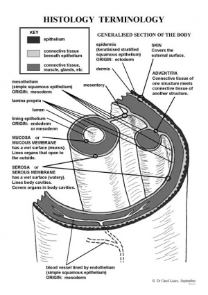
Epithelium forms continuous layers of cells that cover surfaces and line cavities of the body. These cavities include the closed peritoneal, pleural, and pericardial cavities, where the epithelium is called mesothelium, and open organ cavities, i.e. digestive, respiratory, and urogenital organs, which connect with the outside. In addition, epithelium lines the cardiovascular and lymph passageways as endothelium. The parenchymal (secretory) cells of glands (e.g. sweat, salivary) are also epithelium. Epithelial cells are always in close apposition to each other, with a space between membranes of only about 20nm. A small amount of intercellular material, called cement substance (glycosaminoglycan) allows cells to glide over each other and offers only minimal resistance to the migration of leukocytes and other connective tissue cells through the epithelial layers. Epithelial attachment points (junctional complexes) occur between neighboring epithelial cells to hold adjacent cell membranes in close apposition. They also serve as anchoring sites for the fine filaments of the cytoskeleton, which assists in stabilizing the cell shape. Epithelial cells rest on a basal lamina separating them from underlying connective tissue (CT). Epithelium is avascular and for its nutrition depends on diffusion of substances across the basement membrane.
Simple Epithelium
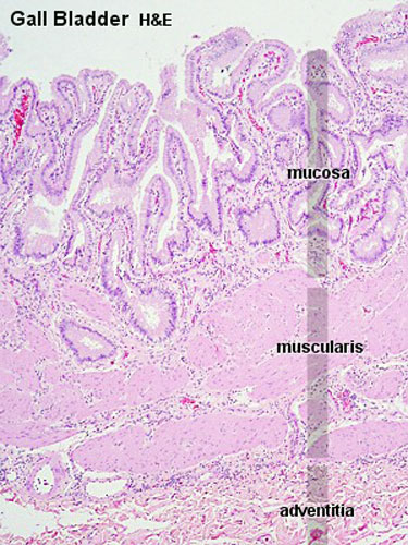
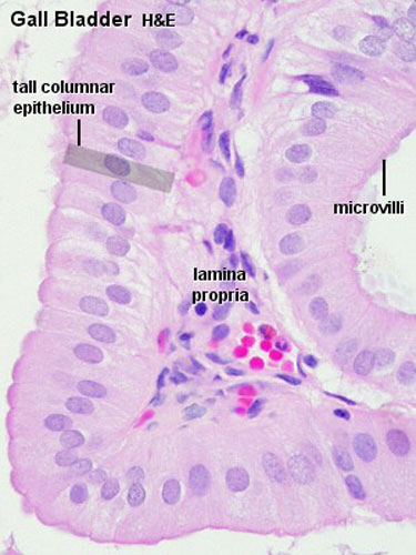
VIRTUAL SLIDE: Gallbladder eg simple epithelium
Stratified Epithelium
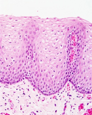
VIRTUAL SLIDE: Oesophagus eg stratified epithelium
Glands
Glands are invaginations of epithelial surfaces that are formed during embryonic development by proliferation of epithelium into the underlying connective tissues to form secretory units. The secretory units, along with their ducts, are the parenchyma of the gland; the stroma of the gland represents the elements of the connective tissue that support the parenchyma. Some glands (endocrine) loose the duct and secrete directly into the blood (e.g. hormones).
Terms
(would need to add a brief description for each term)
- connective tissue
- epithelium
- histology
Glossary Links
- Glossary: A | B | C | D | E | F | G | H | I | J | K | L | M | N | O | P | Q | R | S | T | U | V | W | X | Y | Z | Numbers | Symbols | Term Link
Cite this page: Hill, M.A. (2024, June 2) Embryology Foundations - Histology Cells and Tissues. Retrieved from https://embryology.med.unsw.edu.au/embryology/index.php/Foundations_-_Histology_Cells_and_Tissues
- © Dr Mark Hill 2024, UNSW Embryology ISBN: 978 0 7334 2609 4 - UNSW CRICOS Provider Code No. 00098G


