2011 Lab 3 - Week 3
| 2011 Lab 3: Introduction | Week 3 | Week 4 | Abnormalities | Online Assessment | Group Project |
Folding
Endoderm, mesoderm and ectoderm layers. There are two major folding processes that take place during this time.
- Folding of the whole embryonic disc ventrally, separates the endoderm to form the epithelial lining of the gut. Folding of the embryonic disc occurs ventrally around the notochord, which forms a rod-like region running rostro-caudally in the midline.
- Folding of the ecoderm will form a neural groove, then closing to form a neural tube, separating the neural ectoderm from the embryo surface ectoderm.
Mesoderm Segmentation
Different regions of mesoderm form early intermediate structures.
- Somitogenesis - when part of the mesoderm layer segments during week 3 to form balls of mesoderm called somites. The later migration of cells forms the mesoderm germ layer. An embryonic connective tissue (mesenchyme) which forms nearly all the connective tissues of the body (the head is different). Somitogenesis is when part of this layer segments during week 3 to form balls of mesoderm called somites.
- Intraembryonic coelom - Within the embryonic disc lateral plate mesoderm a space (coelom) forms, it lies within the embryo and so is called the intraembryonic coelom. This single "horseshoe-shaped" space will form the 3 major body cavities: pericardial (around the heart), pleural (around the lungs) and peritoneal (around the GIT and visceral organs).
Ectoderm Segmentation
The central portion of the embryonic disc forms the neural plate, the edge of this plate forms neural crest and the edge forms the epitheium of the skin. This will be covered in week 4.
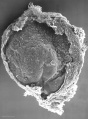
Stage 7
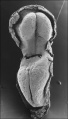
Stage 8
Stage 9


Folding
There are two major folding processes that take place during this time.
- Folding of the ecoderm will form a neural groove, then closing to form a neural tube, separating the neural ectoderm from the embryo surface ectoderm.
- Folding of the whole embryonic disc ventrally, separates the endoderm to form the epithelial lining of the gut. Folding of the embryonic disc occurs ventrally around the notochord, which forms a rod-like region running rostro-caudally in the midline.
![]()
![]()
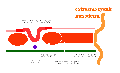
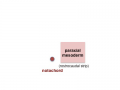
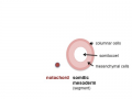
In relation to the notochord:
- Laterally (either side of the notochord) lies mesoderm.
- Rostrally (above the notochord end) lies the buccopharyngeal membrane, above this again is the mesoderm region forming the heart.
- Caudally (below the notochord end) lies the primitive streak (where gastrulation occurred), below this again is the cloacal membrane.
- Dorsally (above the notochord) lies the neural tube then ectoderm.
- Ventrally (beneath the notochord) lies the mesoderm then endoderm.
The ventral endoderm (shown yellow) has grown to line a space called the yolk sac. Folding of the embryonic disc "pinches off" part of this yolk sac forming the first primative GIT.

![]()
Mesoderm
Mesoderm means the "middle layer" and it is from this layer that nearly all the bodies connective tissues are derived. In early mesoderm development a number of transient structures will form and then be lost as tissue structure is patterned and organised. Humans are vertebrates, with a "backbone", and the first mesoderm structure we will see form after the notochord will be somites.
Facts: Week 4, 22 - 23 days, 2 - 3.5 mm, Somite Number 4 - 12
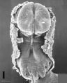
View: This is a dorsal view of the human embryo, the amniotic membrane has been removed. Top embryo is an early stage 10, bottom is late stage 10.
Early stage 10
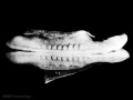
Late stage 10
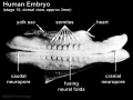
Labeled stage 10


trilaminar embryo
mesoderm regions
somite coelom
neural tube and neural crest


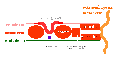
Mesoderm Development
- epiblast -> mesoderm + axial mesoderm (notochord)
- lateral plate + paraxial mesoderm + axial mesoderm
- lateral plate + intermediate mesoderm + somites (body), paraxial mesoderm (head) + axial mesoderm
- somatic mesoderm + intraembryonic coelom + splanchnic mesoderm + intermediate mesoderm + somites (body), paraxial mesoderm (head) + axial mesoderm
Axial Mesoderm

 |
The notochord
Adult - contributes to the nucleus pulposus of the intervertebral disc |
Paraxial Mesoderm
 |
Adult - contributes vertebral column (vertebra and IVD), dermis of the skin, skeletal muscle of body and limbs |
Intermediate Mesoderm
 |
Adult - metanephros forms the kidney |
Lateral Plate Mesoderm
 |
Adult - body and limb connective tissues, gastrointestinal tract (connective tissues, muscle, organs), heart |
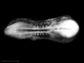
Somite Development

Somite initially forms 2 main components
- ventromedial- sclerotome forms vertebral body and intervertebral disc
- dorsolateral - dermomyotome forms dermis and skeletal muscle
paraxial mesoderm
early somite


Sclerotome
|
Myotome
 |
Forms 2 muscle groups in body and limbs
|
| Development of the sclerotome and myotome components of the somite. |
Dermatome
- connective tissue underlying epidermis
- begins as a dorsal thickening
- spreads throughout the body
Note - Dermatome is the term also used clinically postnatally to describe the region of skin supplied by a single spinal nerve.
Week 2 and 3 Movies
| Implantation | Mesoderm | Chorionic Cavity | Amniotic Cavity | Week 3 |
Embryo Stages and Events
| Day | Stage | Event |
| Stage 7 |  | |
| Stage 8 |  | |
 | ||
| Stage 9 | 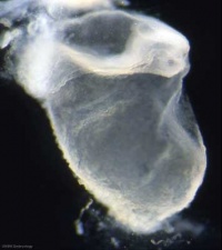 Musculoskeletal System Development somitogenesis - first somites form and continue to be added in sequence caudally Musculoskeletal System Development somitogenesis - first somites form and continue to be added in sequence caudally
Neural System Development - three main divisions of the brain, which are not cerebral vesicles, can be distinguished while the neural groove is still completely open
| |
| Cardiovascular System Development cardiogenesis - week 3 begins as paired heart tubes. |
| 2011 Lab 3: Introduction | Week 3 | Week 4 | Abnormalities | Online Assessment | Group Project |