File:Gray0015.jpg
From Embryology
Size of this preview: 703 × 599 pixels.
Original file (800 × 682 pixels, file size: 111 KB, MIME type: image/jpeg)
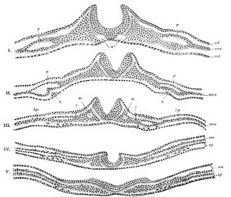
Neural Groove
A series of transverse sections through an embryo of the dog. (After Bonnet.)
The series shows the uprising of the neural folds to form the neural canal.
Section I is the most anterior. In V the neural plate is spread out nearly flat.
In III, IV, and V the scattered cells represented between the entoderm and splanchnic layer of mesoderm
are the vasoformative cells which give origin in front, according to Bonnet, to the heart tubes,
h; l.p. Lateral plate still undivided in I, II, and III; in IV and V split into somatic (sm) and splanchnic (sp) layers
of mesoderm.
Legend
- a. Aortæ.
- c. Intermediate cell mass.
- ect. Ectoderm.
- ent. Entoderm. (endoderm)
- h. Rudiments of endothelial heart tubes.
- mes. Mesoderm.
- p. Pericardium.
- so. Primitive segment. (somite)
File history
Click on a date/time to view the file as it appeared at that time.
| Date/Time | Thumbnail | Dimensions | User | Comment | |
|---|---|---|---|---|---|
| current | 13:45, 23 October 2010 | | 800 × 682 (111 KB) | S8600021 (talk | contribs) | ==Neural Groove== A series of transverse sections through an embryo of the dog. (After Bonnet.) The series shows the uprising of the neural folds to form the neural canal. Section I is the most anterior. In V the neural plate is spread out nearly fla |
You cannot overwrite this file.
File usage
The following 2 pages use this file: