File:Thyroid-development-cartoon.jpg
Thyroid-development-cartoon.jpg (600 × 541 pixels, file size: 32 KB, MIME type: image/jpeg)
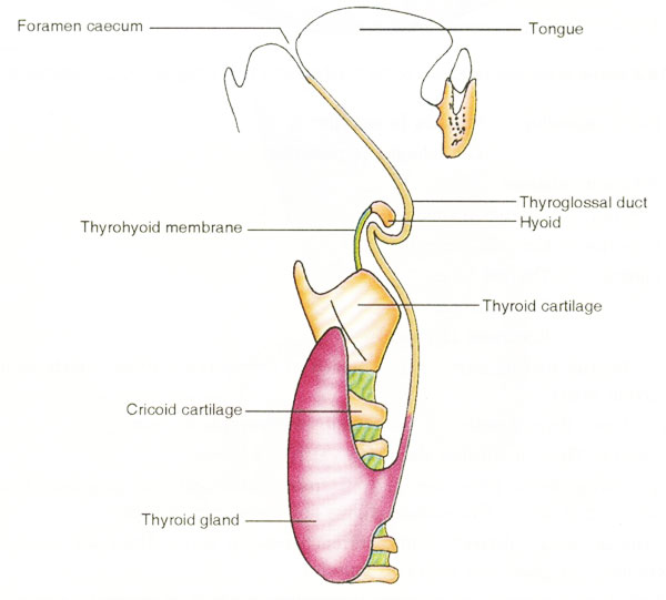
Showing the development of thyroid gland. The thyroid gland begins to develop as a median thickening of endoderm on the floor of the pharynx between the first and second pharyngeal pouches. This area later invaginates to form the median diverticulum, which appears in the later half of the fourth week. This thyroid diverticulum grows further, becoming a solid cellular cord called the thyroglossal duct. The duct grows caudally and bifurcates to give rise to the thyroid lobes and the isthmus.
Original file name: Figure 4 http://www.ncbi.nlm.nih.gov/pmc/articles/PMC2827060/figure/F4/
Reference
<pubmed>20181171</pubmed>| PMC2827060
This is an Open Access article distributed under the terms of the Creative Commons Attribution License (http://creativecommons.org/licenses/by/3.0), which permits unrestricted use, distribution, and reproduction in any medium, provided the original work is properly cited.
File history
Click on a date/time to view the file as it appeared at that time.
| Date/Time | Thumbnail | Dimensions | User | Comment | |
|---|---|---|---|---|---|
| current | 23:20, 5 October 2010 | | 600 × 541 (32 KB) | S8600021 (talk | contribs) | Showing the development of thyroid gland. The thyroid gland begins to develop as a median thickening of endoderm on the floor of the pharynx between the first and second pharyngeal pouches. This area later invaginates to form the median diverticulum, whi |
You cannot overwrite this file.
File usage
The following 7 pages use this file: