File:Tooth stage lamina.jpg
Tooth_stage_lamina.jpg (430 × 352 pixels, file size: 22 KB, MIME type: image/jpeg)
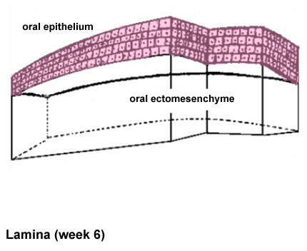
Stages in Tooth Development
Pre-patterned oral ectoderm is in close contact with cranial, neural crest ectomesenchyme. At this stage (mouse E10) the odontogenic potential resides in the epithelium.
Related Images: lamina | placode stage | bud stage | cap stage | bell stage
Adapted from original file name: Ijbsv05p0226g04.jpg http://www.pubmedcentral.nih.gov/articlerender.fcgi?artid=2651620&rendertype=figure&id=F4
Reference
<pubmed>19266065</pubmed>| PMCID: PMC2651620
Int J Biol Sci. 2009; 5(3): 226–243. Published online 2009 February 24.
Copyright © Ivyspring International Publisher. This is an open-access article distributed under the terms of the Creative Commons License (http://creativecommons.org/licenses/by-nc-nd/3.0/). Reproduction is permitted for personal, noncommercial use, provided that the article is in whole, unmodified, and properly cited.
File history
Click on a date/time to view the file as it appeared at that time.
| Date/Time | Thumbnail | Dimensions | User | Comment | |
|---|---|---|---|---|---|
| current | 10:48, 21 September 2010 | | 430 × 352 (22 KB) | S8600021 (talk | contribs) | ==Stages in Tooth Development== Pre-patterned oral ectoderm is in close contact with cranial, neural crest ectomesenchyme. At this stage (mouse E10) the odontogenic potential resides in the epithelium. Adapted from original file name: Ijbsv05p0226g04 |
You cannot overwrite this file.