File:Cullen1916 fig33.jpg

Original file (1,154 × 2,052 pixels, file size: 422 KB, MIME type: image/jpeg)
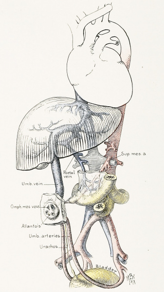
Fig. 33. A Diagrammatic Representation of the Umbilical Region of a Fetus at Term
An exceptional feature of the picture is the persistence of the omphalomesenteric structures in the form of a well defined vascular Meckel's diverticulum.
Note the relationship between the omphalomesenteric vessels, the superior mesenteric artery, and the portal vein. In the cord the vitelline vessels still persist in the organized exocoelom.
At birth all trace of the omphalomesenteric structure has usually disappeared, but the allantoic cord often persists, and is seen between and below the umbilical arteries. It is continuous with the urachus, which may have a number of spindle-shaped dilatations down to the bladder. These may or may not have lumina.
| Historic Disclaimer - information about historic embryology pages |
|---|
 |
- Figure Links: 1 Human embryo 0.7 mm | 2 Human embryo 1.7 mm | 3 Human embryo 2.5 mm | 4 Human embryo 3.5 mm | 5 Human embryo 5 mm | 6 Human embryo 7 mm | 7 Human embryo 7 mm | 8 Human embryo 10 mm | 9 Human embryo 12.5 mm | 10 Human embryo 10 mm | 11 Human embryo 23 mm | 12 Human embryo 3 cm | 13 Human embryo 4.5 cm sagittal | 14 Human Embryo 4.5 cm | 15 Human Embryo 5.2 cm | 16 Human Embryo 6.5 cm | 17 Human Embryo 7.5 cm | 18 Human Embryo 9 cm | 19 Human Embryo 10 cm | 20 Human Embryo 12 cm | 21 Human Embryo 12 cm | 22 Human Embryo 12 cm | 23 Human Embryo 12 cm Cord | 28 Fetus Five Months | 30 Ventral Heria | 31 Human Embryo 5.5 cm | 32 Term Human | 33 Term Human | [[Figures
Reference
Cullen TS. Embryology, anatomy, and diseases of the umbilicus together with diseases of the urachus. (1916) W. B. Saunders Company, Philadelphia And London.
Cite this page: Hill, M.A. (2024, June 10) Embryology Cullen1916 fig33.jpg. Retrieved from https://embryology.med.unsw.edu.au/embryology/index.php/File:Cullen1916_fig33.jpg
- © Dr Mark Hill 2024, UNSW Embryology ISBN: 978 0 7334 2609 4 - UNSW CRICOS Provider Code No. 00098G
File history
Click on a date/time to view the file as it appeared at that time.
| Date/Time | Thumbnail | Dimensions | User | Comment | |
|---|---|---|---|---|---|
| current | 13:46, 28 October 2018 | | 1,154 × 2,052 (422 KB) | Z8600021 (talk | contribs) | |
| 13:41, 28 October 2018 | 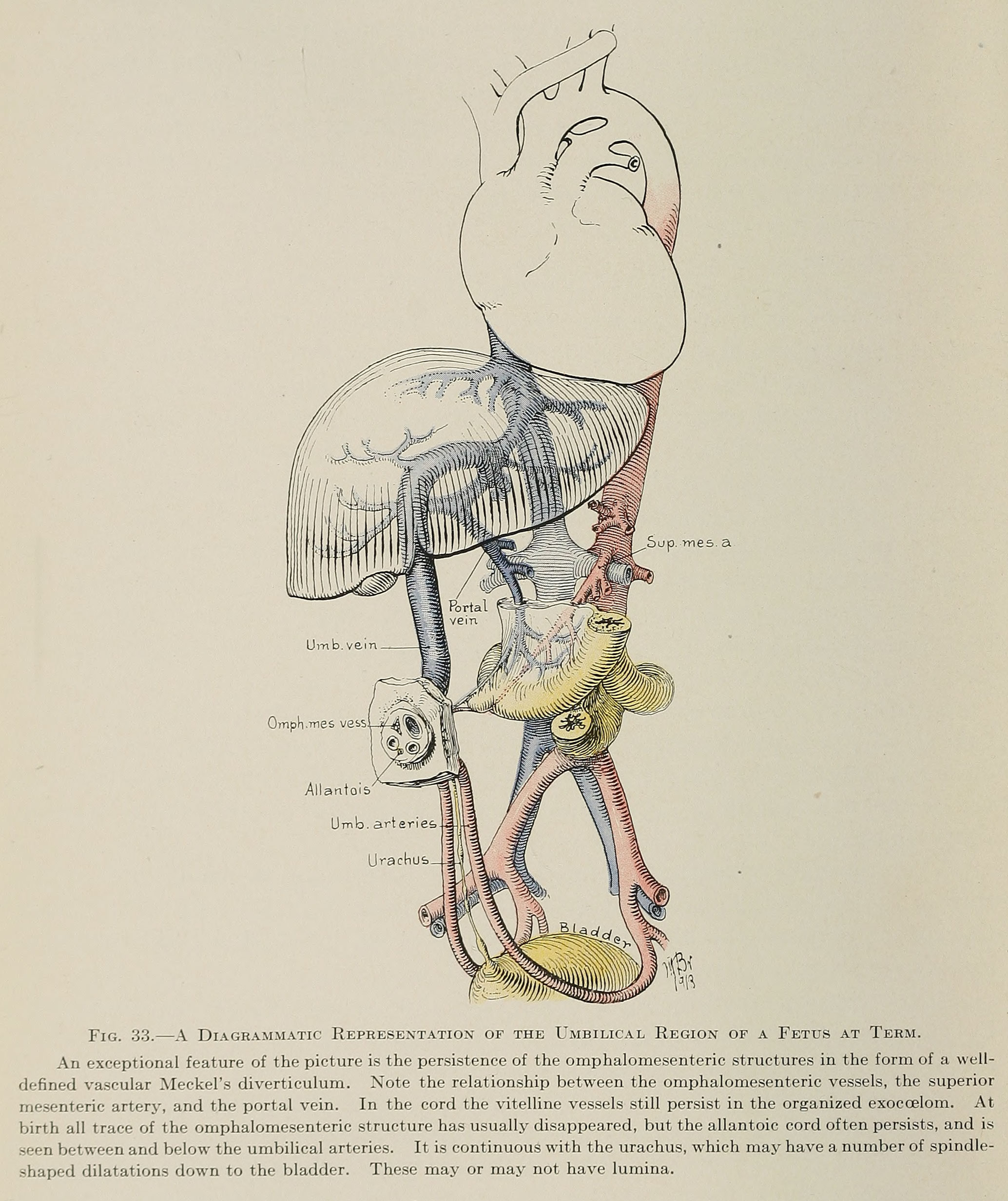 | 2,068 × 2,466 (690 KB) | Z8600021 (talk | contribs) | ==Fig. 33. A Diagrammatic Representation of the Umbilical Region of a Fetus at Term== An exceptional feature of the picture is the persistence of the omphalomesenteric structures in the form of a welldefined vascular Meckel's diverticulum. Note the re... |
You cannot overwrite this file.
File usage
The following 3 pages use this file: