File:Altschule1930 fig01-02.jpg
From Embryology

Size of this preview: 484 × 599 pixels. Other resolution: 1,000 × 1,238 pixels.
Original file (1,000 × 1,238 pixels, file size: 175 KB, MIME type: image/jpeg)
Fig. 1 Reconstruction of part of the right mesonephros of a human embryo of 36 mm.
Fig. 2 Reconstruction of part of the right mesonephros of a human embryo of 31 mm.
Reference
Altschule MD. The changes in the mesonephric tubules of human embryos ten to twelve weeks old. (1930) Anat. Rec. 46(1): 81-91.
Cite this page: Hill, M.A. (2024, June 5) Embryology Altschule1930 fig01-02.jpg. Retrieved from https://embryology.med.unsw.edu.au/embryology/index.php/File:Altschule1930_fig01-02.jpg
- © Dr Mark Hill 2024, UNSW Embryology ISBN: 978 0 7334 2609 4 - UNSW CRICOS Provider Code No. 00098G
File history
Click on a date/time to view the file as it appeared at that time.
| Date/Time | Thumbnail | Dimensions | User | Comment | |
|---|---|---|---|---|---|
| current | 13:37, 26 February 2017 | | 1,000 × 1,238 (175 KB) | Z8600021 (talk | contribs) | |
| 13:35, 26 February 2017 | 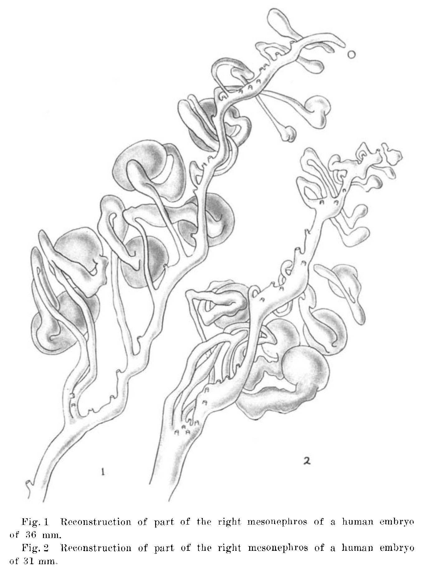 | 1,358 × 1,833 (235 KB) | Z8600021 (talk | contribs) |
You cannot overwrite this file.
File usage
The following page uses this file: