File:Gage1905-fig01.jpg
Original file (1,183 × 1,500 pixels, file size: 301 KB, MIME type: image/jpeg)
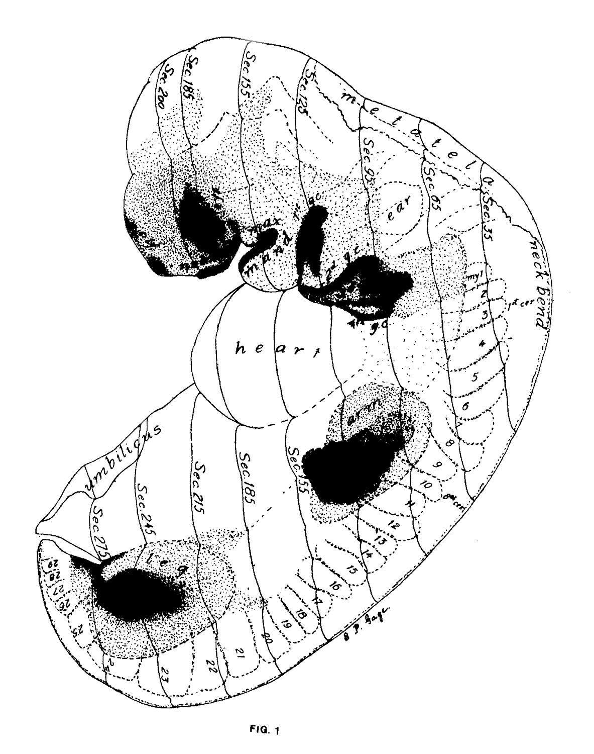
Fig. 1. View of the left side of the model
Compare with figures of this embryo in articles by Mall 1, 5.
It shows: The head comparatively small in diameter but great in length, and forming at the neck-bend an angle of 65° with the body; the position of the neuropore; the eye and ear scarcely apparent as external features; the "prominent heart, limb buds, and tail; the umbilicus turning to the right (cf. Fig. 5); the wide undeveloped mouth and small maxillary process; the crowding of the 2d, 3d, and 4th clefts into the precervical sinus; and 29 myotomes, the 3d being noted as the 1st cervical.
The density of the stippling on Fig. 1 indicates the relative thickness of the epithelium (see above, External Form).
The topographic lines show the direction of the sections, the numbers upon them indicate the corresponding sections of the eries. The following figures have either topographic lines or the section number at which they are cut, and hence can be located with reference to Fig. 1.
Reference
Gage SP. A three weeks' human embryo, with especial reference to the brain and nephric system. (1905) Amer. J Anat. 4: 409-443.
Cite this page: Hill, M.A. (2024, June 2) Embryology Gage1905-fig01.jpg. Retrieved from https://embryology.med.unsw.edu.au/embryology/index.php/File:Gage1905-fig01.jpg
- © Dr Mark Hill 2024, UNSW Embryology ISBN: 978 0 7334 2609 4 - UNSW CRICOS Provider Code No. 00098G
File history
Click on a date/time to view the file as it appeared at that time.
| Date/Time | Thumbnail | Dimensions | User | Comment | |
|---|---|---|---|---|---|
| current | 12:59, 18 August 2016 | | 1,183 × 1,500 (301 KB) | Z8600021 (talk | contribs) | ===Reference=== {{Ref-Gage1905}} |
You cannot overwrite this file.
File usage
The following page uses this file: