File:Stage11 sem5c.jpg

Stage11_sem5c.jpg (245 × 400 pixels, file size: 14 KB, MIME type: image/jpeg)
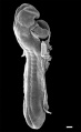
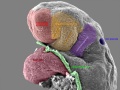
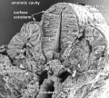
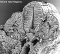
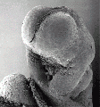
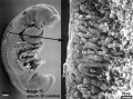
Human Embryo
Carnegie stage 11 24 days, 13 somite pairs
Facts: Week 4, 23 - 26 days, 2.5 - 4.5 mm, Somite Number 13 - 20
View: This is a scanning EM of the embryo dorsal view showing the neural tube closing with open neuropores and the paired somites visible through the thin ectoderm.
Features: surface ectoderm, neural tube, cranial (anterior) neuropore, caudal (posterior) neuropore, somites, heart, cut edge of amnion
Stage11_sem5c.jpg
Original file name: Stage11day24somite13-dorsal-sem5-1000px.jpg
Image version links: Large 1000px | 800px |
Medium 600px | Small 400px
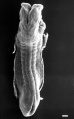
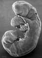
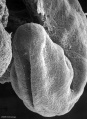
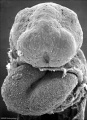
- Stage 11 SEM Images: dorsolateral whole embryo | dorsal embryo | lateral embryo | lateral head | lateral head with overlay | embryo cross-section | ventrolateral head | ventrolateral head with overlay | ventral head | buccopharyngeal membrane | neural crest | posterior neuropore | anterior neuropore | Carnegie stage 11
- Human Embryo (stage 11)
dorsolateral whole embryo
dorsal embryo
lateral embryo
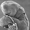
lateral head
lateral head with overlay

embryo cross-section
embryo cross-section label
neural cross-section label

ventrolateral head
ventrolateral head with overlay

ventral head

buccopharyngeal membrane
neural crest
posterior neuropore
anterior neuropore


Image Source: Scanning electron micrographs of the Carnegie stages of the early human embryos are reproduced with the permission of Prof Kathy Sulik, from embryos collected by Dr. Vekemans and Tania Attié-Bitach. Images are for educational purposes only and cannot be reproduced electronically or in writing without permission.
- Carnegie Stages: 1 | 2 | 3 | 4 | 5 | 6 | 7 | 8 | 9 | 10 | 11 | 12 | 13 | 14 | 15 | 16 | 17 | 18 | 19 | 20 | 21 | 22 | 23 | About Stages | Timeline
Cite this page: Hill, M.A. (2024, May 23) Embryology Stage11 sem5c.jpg. Retrieved from https://embryology.med.unsw.edu.au/embryology/index.php/File:Stage11_sem5c.jpg
- © Dr Mark Hill 2024, UNSW Embryology ISBN: 978 0 7334 2609 4 - UNSW CRICOS Provider Code No. 00098G
File history
Click on a date/time to view the file as it appeared at that time.
| Date/Time | Thumbnail | Dimensions | User | Comment | |
|---|---|---|---|---|---|
| current | 01:20, 6 May 2010 | | 245 × 400 (14 KB) | S8600021 (talk | contribs) | '''Human Embryo''' Carnegie stage 11 24 days, 13 somite pairs Facts: Week 4, 23 - 26 days, 2.5 - 4.5 mm, Somite Number 13 - 20 View: This is a scanning EM of the embryo dorsal view showing the neural tube closing with open neuropores and the paired som |
You cannot overwrite this file.
File usage
The following 6 pages use this file: