File:Liver structure cartoon.jpg
Original file (1,000 × 451 pixels, file size: 78 KB, MIME type: image/jpeg)
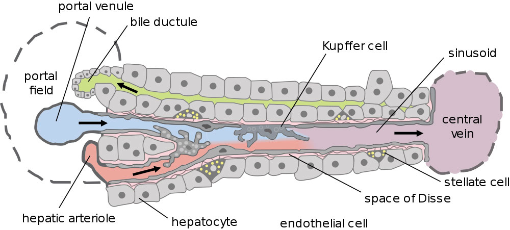
Liver Structure
This cartoon shows the portal venue to central vein flow within the liver subunit structure.
- Dual blood supply of the liver merges upon entry into the liver lobule at the portal field.
- branches of the portal vein
- branches of the hepatic artery
- Blood flows along the sinusoid and exits at the central vein.
- Bile flows in the opposite direction.
Kupffer cells - stellate macrophage cells named after Karl Wilhelm von Kupffer (1829 - 1902) a German anatomist who originally identified these cells.
- Links: Liver Development | Image- Model of Plasmodium Sporozoite Infection of the Mammalian Liver | Image- Liver structure cartoon
Reference
<pubmed>15901208</pubmed>| PLoS Biol
Copyright
© 2005 Frevert et al. This is an open-access article distributed under the terms of the Creative Commons Attribution License, which permits unrestricted use, distribution, and reproduction in any medium, provided the original work is properly cited.
http://journals.plos.org/plosbiology/article?id=10.1371/journal.pbio.0030192
Cite this page: Hill, M.A. (2024, May 23) Embryology Liver structure cartoon.jpg. Retrieved from https://embryology.med.unsw.edu.au/embryology/index.php/File:Liver_structure_cartoon.jpg
- © Dr Mark Hill 2024, UNSW Embryology ISBN: 978 0 7334 2609 4 - UNSW CRICOS Provider Code No. 00098G
File history
Click on a date/time to view the file as it appeared at that time.
| Date/Time | Thumbnail | Dimensions | User | Comment | |
|---|---|---|---|---|---|
| current | 09:15, 4 May 2011 | | 1,000 × 451 (78 KB) | S8600021 (talk | contribs) | ==Liver Structure== Dual blood supply of the liver merges upon entry into the liver lobule at the portal field. # branches of the portal vein # branches of the hepatic artery The blood flows along the sinusoid and exits at the central vein. (text mod |
You cannot overwrite this file.
File usage
The following 10 pages use this file:
- 2011 Lab 5 - Late Embryo
- ANAT2241 Liver, Gallbladder, and Pancreas
- ANAT2341 Lab 5 - Late Embryo
- BGDB Gastrointestinal - Activity 3
- BGDB Gastrointestinal - Late Embryo
- BGD Lecture - Gastrointestinal System Development
- Gastrointestinal Tract - Liver Development
- Gastrointestinal Tract - Liver Histology
- Lecture - Gastrointestinal Development 2013
- Template talk:Embryonic Liver Timeline Table