File:Liver structure cartoon.jpg
Original file (1,000 × 451 pixels, file size: 78 KB, MIME type: image/jpeg)
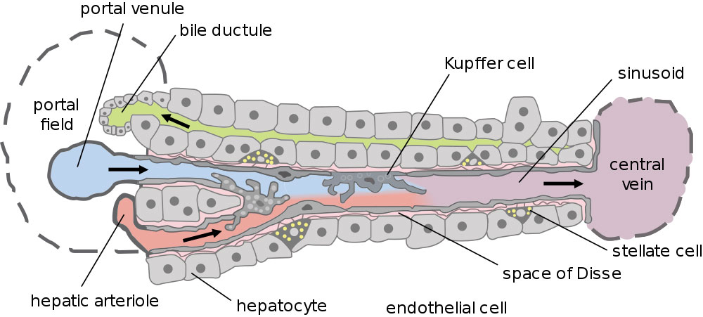
Liver Structure
This cartoon shows the portal venue to central vein flow within the liver subunit structure.
- Dual blood supply of the liver merges upon entry into the liver lobule at the portal field.
- branches of the portal vein
- branches of the hepatic artery
- Blood flows along the sinusoid and exits at the central vein.
- Bile flows in the opposite direction.
Kupffer cells - stellate macrophage cells named after Karl Wilhelm von Kupffer (1829 - 1902) a German anatomist who originally identified these cells.
- Links: Liver Development | Image- Model of Plasmodium Sporozoite Infection of the Mammalian Liver | Image- Liver structure cartoon
Reference
<pubmed>15901208</pubmed>| PLoS Biol
Copyright
© 2005 Frevert et al. This is an open-access article distributed under the terms of the Creative Commons Attribution License, which permits unrestricted use, distribution, and reproduction in any medium, provided the original work is properly cited.
Cite this page: Hill, M.A. (2024, May 23) Embryology Liver structure cartoon.jpg. Retrieved from https://embryology.med.unsw.edu.au/embryology/index.php/File:Liver_structure_cartoon.jpg
- © Dr Mark Hill 2024, UNSW Embryology ISBN: 978 0 7334 2609 4 - UNSW CRICOS Provider Code No. 00098G
File history
Click on a date/time to view the file as it appeared at that time.
| Date/Time | Thumbnail | Dimensions | User | Comment | |
|---|---|---|---|---|---|
| current | 09:15, 4 May 2011 | | 1,000 × 451 (78 KB) | S8600021 (talk | contribs) | ==Liver Structure== Dual blood supply of the liver merges upon entry into the liver lobule at the portal field. # branches of the portal vein # branches of the hepatic artery The blood flows along the sinusoid and exits at the central vein. (text mod |
You cannot overwrite this file.
File usage
The following 10 pages use this file:
- 2011 Lab 5 - Late Embryo
- ANAT2241 Liver, Gallbladder, and Pancreas
- ANAT2341 Lab 5 - Late Embryo
- BGDB Gastrointestinal - Activity 3
- BGDB Gastrointestinal - Late Embryo
- BGD Lecture - Gastrointestinal System Development
- Gastrointestinal Tract - Liver Development
- Gastrointestinal Tract - Liver Histology
- Lecture - Gastrointestinal Development 2013
- Template talk:Embryonic Liver Timeline Table