File:Meyer1914 fig26.jpg
From Embryology
Size of this preview: 635 × 599 pixels. Other resolution: 679 × 641 pixels.
Original file (679 × 641 pixels, file size: 120 KB, MIME type: image/jpeg)
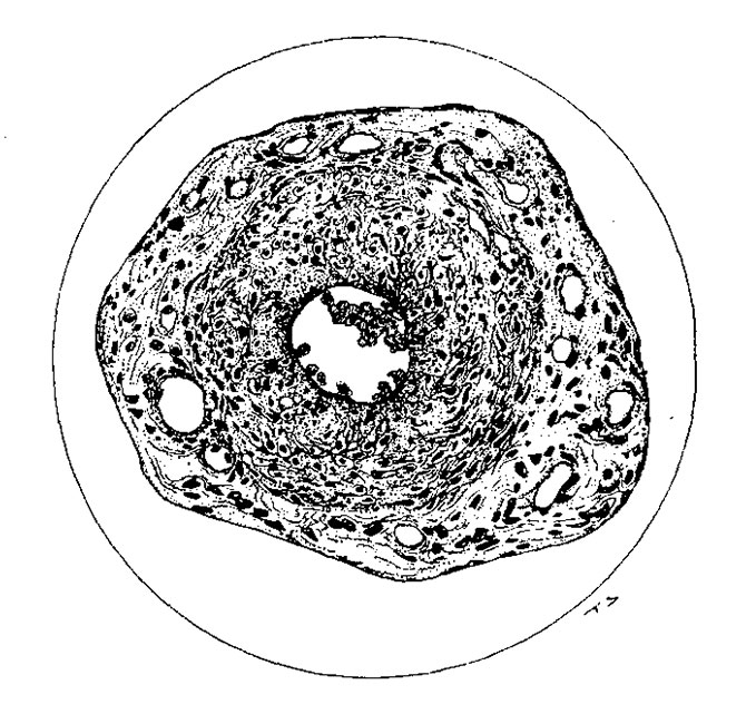
Fig. 26. Omphalomesenteric vessel from a pup 91 hours old. The arrangement of the inner portion of the wall suggests that of the original vessel. The lumen contains well-preserved erythrocytes. The outer lighter portion is composed of very loose fibrous connective tissue. The endothelium is intact in this portion. X275.
File history
Click on a date/time to view the file as it appeared at that time.
| Date/Time | Thumbnail | Dimensions | User | Comment | |
|---|---|---|---|---|---|
| current | 21:08, 3 November 2015 | | 679 × 641 (120 KB) | Z8600021 (talk | contribs) | |
| 21:08, 3 November 2015 | 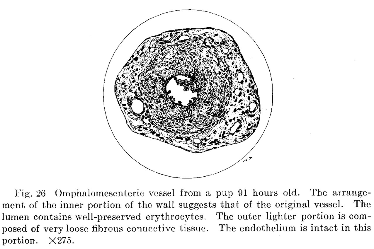 | 1,276 × 850 (194 KB) | Z8600021 (talk | contribs) | Fig. 26. Omphalomesenteric vessel from a pup 91 hours old. The arrangement of the inner portion of the wall suggests that of the original vessel. The lumen contains well-preserved erythrocytes. The outer lighter portion is composed of very loose fibrou... |
You cannot overwrite this file.
File usage
The following page uses this file: