File:Hair follicle development.jpg
Original file (800 × 663 pixels, file size: 191 KB, MIME type: image/jpeg)
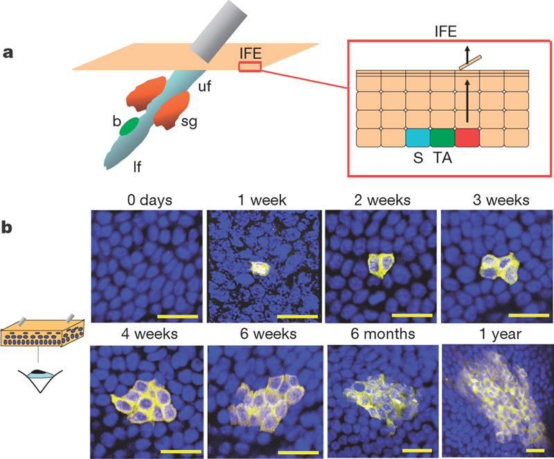
Organization of the epidermis.
Hair follicles contain stem cells located in the bulge (b, green), with the potential to generate lower hair follicle (lf), sebaceous gland (sg, orange) upper follicle (uf) and interfollicular epidermis (IFE, beige). The schematic shows the organization of keratinocytes in the IFE, as proposed by the stem/TA cell hypothesis. The basal layer comprises stem cells (S, blue), transit amplifying cells (TA, dark green), and post-mitotic basal cells (red), which migrate out of the basal layer as they differentiate (arrows).
Original file name: Nature05574-f1.2.jpg
A single type of progenitor cell maintains normal epidermis. Clayton E, Doupe DP, Klein AM, Winton DJ, Simons BD, Jones PH. Nature. 2007 Mar 8;446(7132):185-9. PMID: 17330052
Reprinted by permission from Macmillan Publishers Ltd: Nature. 2007 Mar 8;446(7132):185-9, copyright (2007)
File history
Click on a date/time to view the file as it appeared at that time.
| Date/Time | Thumbnail | Dimensions | User | Comment | |
|---|---|---|---|---|---|
| current | 00:37, 22 April 2010 | | 800 × 663 (191 KB) | S8600021 (talk | contribs) | Organization of the epidermis. Hair follicles contain stem cells located in the bulge (b, green), with the potential to generate lower hair follicle (lf), sebaceous gland (sg, orange) upper follicle (uf) and interfollicular epidermis (IFE, beige). The s |
You cannot overwrite this file.
File usage
The following 2 pages use this file: