File:Stricht-plate03.jpg
Original file (1,023 × 1,260 pixels, file size: 230 KB, MIME type: image/jpeg)
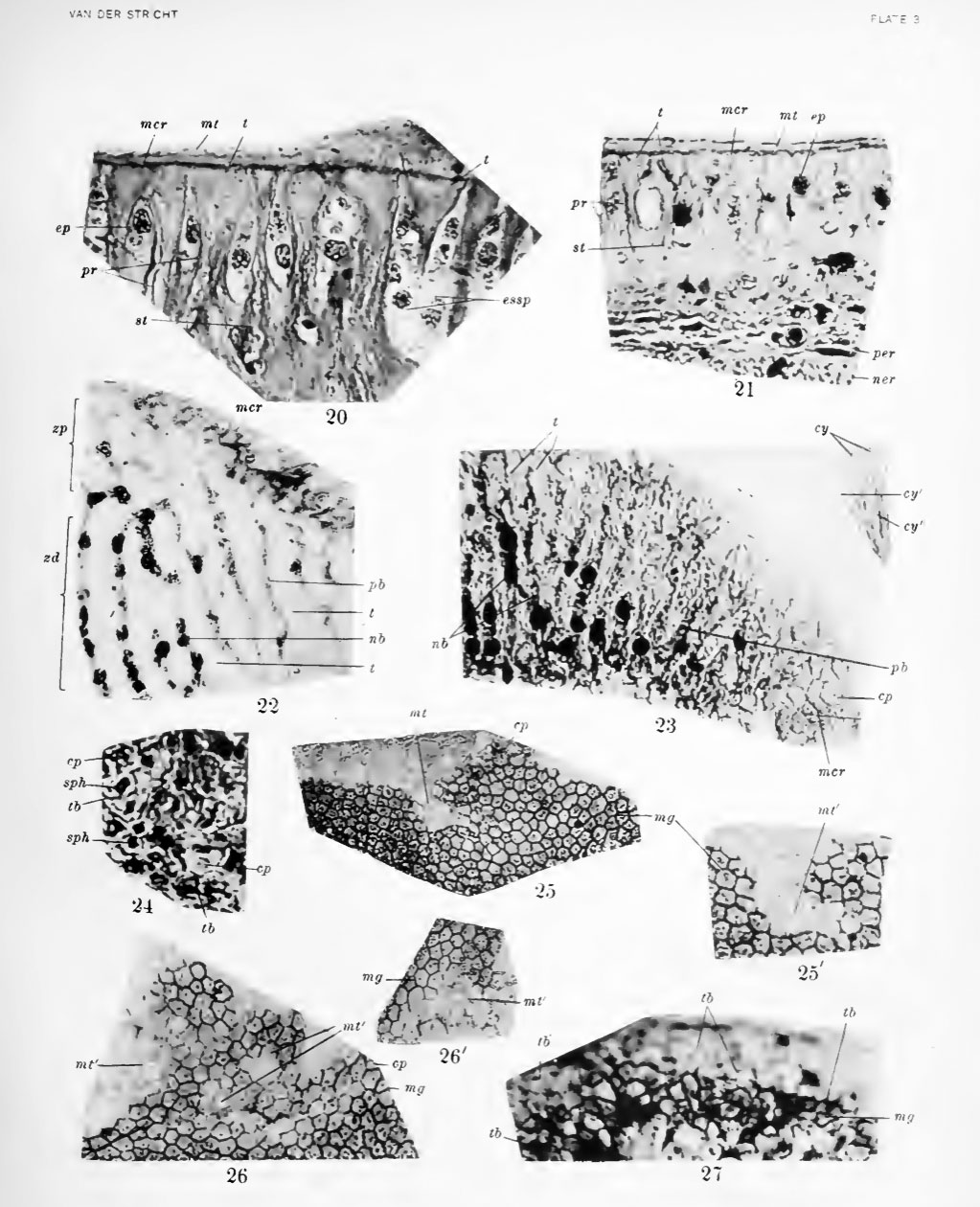
Plate 3
20. Photograph of section vertical to surface of crista spiralis. Pig embryo 190 mm. Bouin's fluid; Mallory's stain.
21. Photograph of section vertical to surface of crista spiralis. Young dog, age 4 months. Trichloracetic acid: iron hematoxylin, Congo red, light green.
22. Photograph of section tangential to surface of crista spiralis. Pig embryo 190 mm. Trichloracetic acid; iron hematoxylin, Congo red.
23. Photograph of section tangential to surface of crista spiralis in adult bat (Vesperlilw fuscus). Zenker's fluid, iron hematoxylin, Congo red.
24. Photograph of section tangential to surface of crista spiralis. Young dog. Bouin's fluid; iron hematoxylin; Congo red.
25, 25', 26, 26'. Photographs of sections tangential to surface of greater ridge. New-born dog. Trichloracetic acid; iron hematoxylin, Congo red.
27. Photograph of section tangential to surface of greater ridge. Pig embryo 93.5 mm. Fixation by uranium nitrate method of Ramon y Cajal.
File history
Click on a date/time to view the file as it appeared at that time.
| Date/Time | Thumbnail | Dimensions | User | Comment | |
|---|---|---|---|---|---|
| current | 08:56, 7 April 2011 | | 1,023 × 1,260 (230 KB) | S8600021 (talk | contribs) | ==Plate 3== 20. Photograph of section vertical to surface of crista spiralis. Pig embryo 190 mm. Bouin's fluid; Mallory's stain. 21. Photograph of section vertical to surface of crista spiralis. Young dog, age 4 months. Trichloracetic acid: iron hemato |
You cannot overwrite this file.
File usage
The following page uses this file: