File:Gray0379.jpg
Gray0379.jpg (713 × 600 pixels, file size: 114 KB, MIME type: image/jpeg)
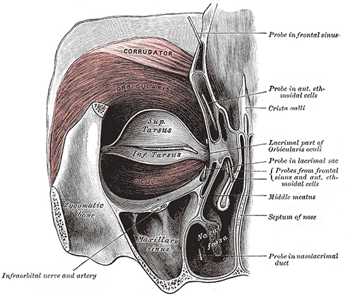
Fig. 379. Left orbicularis oculi
(seen from behind)
The Orbicularis oculi (Orbicularis palpebrarum) (Fig. 379) arises from the nasal part of the frontal bone, from the frontal process of the maxilla in front of the lacrimal groove, and from the anterior surface and borders of a short fibrous band, the medial palpebral ligament. From this origin, the fibers are directed lateralward, forming a broad and thin layer, which occupies the eyelids or palpebræ, surrounds the circumference of the orbit, and spreads over the temple, and downward on the cheek. The palpebral portion of the muscle is thin and pale; it arises from the bifurcation of the medial palpebral ligament, forms a series of concentric curves, and is inserted into the lateral palpebral raphé. The orbital portion is thicker and of a reddish color; its fibers form a complete ellipse without interruption at the lateral palpebral commissure; the upper fibers of this portion blend with the Frontalis and Corrugator. The lacrimal part (Tensor tarsi) is a small, thin muscle, about 6 mm. in breadth and 12 mm. in length, situated behind the medial palpebral ligament and lacrimal sac (Fig. 379). It arises from the posterior crest and adjacent part of the orbital surface of the lacrimal bone, and passing behind the lacrimal sac, divides into two slips, upper and lower, which are inserted into the superior and inferior tarsi medial to the puncta lacrimalia; occasionally it is very indistinct.
- Gray's Images: Development | Lymphatic | Neural | Vision | Hearing | Somatosensory | Integumentary | Respiratory | Gastrointestinal | Urogenital | Endocrine | Surface Anatomy | iBook | Historic Disclaimer
| Historic Disclaimer - information about historic embryology pages |
|---|
| Pages where the terms "Historic" (textbooks, papers, people, recommendations) appear on this site, and sections within pages where this disclaimer appears, indicate that the content and scientific understanding are specific to the time of publication. This means that while some scientific descriptions are still accurate, the terminology and interpretation of the developmental mechanisms reflect the understanding at the time of original publication and those of the preceding periods, these terms, interpretations and recommendations may not reflect our current scientific understanding. (More? Embryology History | Historic Embryology Papers) |
| iBook - Gray's Embryology | |
|---|---|

|
|
Reference
Gray H. Anatomy of the human body. (1918) Philadelphia: Lea & Febiger.
Cite this page: Hill, M.A. (2024, May 29) Embryology Gray0379.jpg. Retrieved from https://embryology.med.unsw.edu.au/embryology/index.php/File:Gray0379.jpg
- © Dr Mark Hill 2024, UNSW Embryology ISBN: 978 0 7334 2609 4 - UNSW CRICOS Provider Code No. 00098G
File history
Click on a date/time to view the file as it appeared at that time.
| Date/Time | Thumbnail | Dimensions | User | Comment | |
|---|---|---|---|---|---|
| current | 07:36, 11 May 2014 |  | 713 × 600 (114 KB) | Z8600021 (talk | contribs) | ==Fig. 379. Left orbicularis oculi== (seen from behind) The Orbicularis oculi (Orbicularis palpebrarum) (Fig. 379) arises from the nasal part of the frontal bone, from the frontal process of the maxilla in front of the lacrimal groove, and from the a... |
You cannot overwrite this file.
File usage
The following 2 pages use this file:

