File:Keibel1900 plate01.jpg
Original file (1,952 × 2,451 pixels, file size: 519 KB, MIME type: image/jpeg)
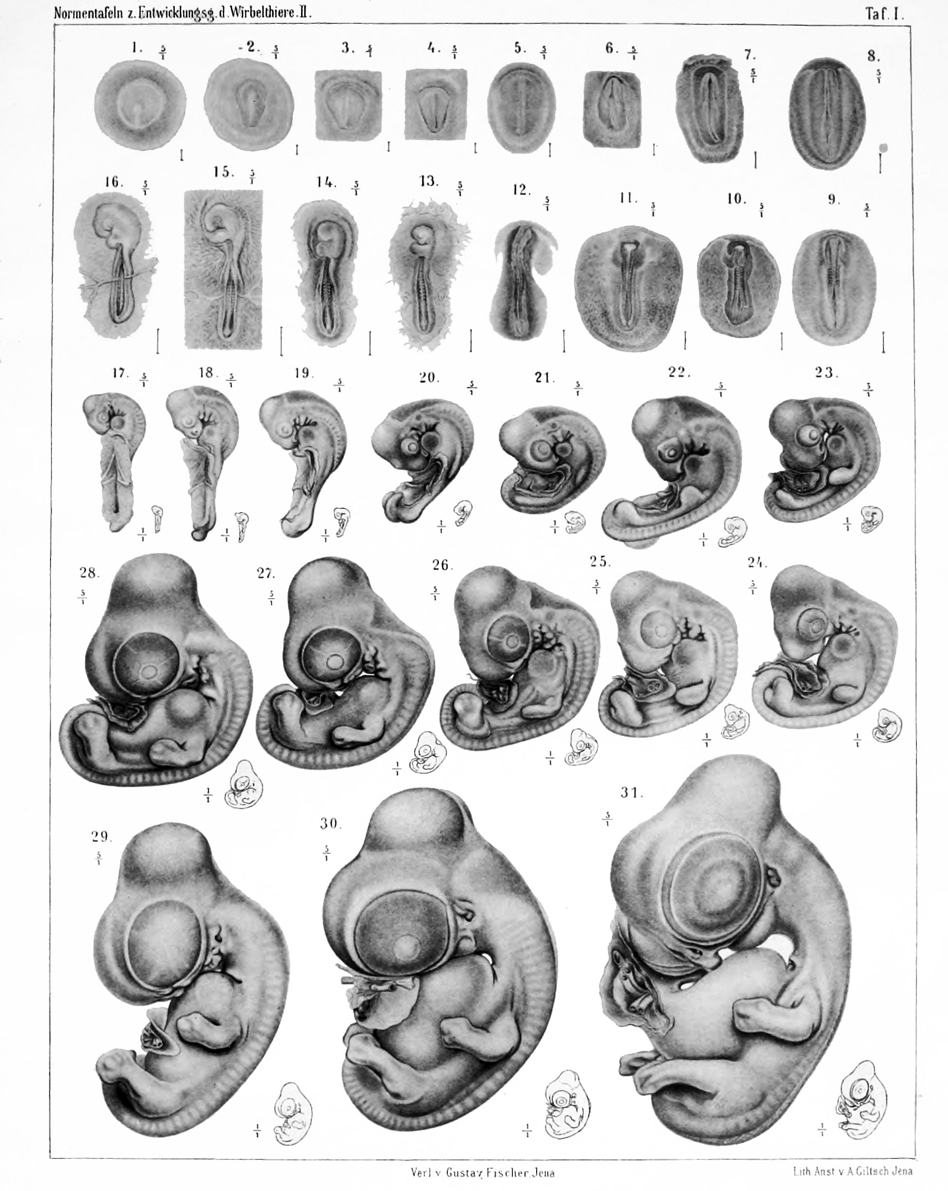
Plate 1
Figure legends text shown below is a modified online translation. Original German text.
| Translation Request |
|---|
 If you would like to help with the online translation into English please contact me. |
Figure 1
(S.N. 452) The germ shown as Figure 1 is taken from a 9 hours embryonated egg fixed with sublimate-acetic acid. The whole seed is approximately circular, its diameter is 4.1 mm. The same in the middle is the zona pellucida, which is near the circular shape, but narrows somewhat towards the rear. Its length is 2.3 mm, and its maximum width 2.15 mm. The primitive streak begins about the middle of the zona pellucida with a button-shaped widening and gradually disappears towards the rear end of the zona pellucida, here a considerable width reaching. Its length is 1.2 mm.
The series shows an early stage of mesoderm formation, the mesoderm does not exceed even the scope of the area pellucida. The primitive streak is already embodied in the typical district front than in the rear. A clear head extension does not exist, nor any traces of blood formation.
Figure 2
(S.N. 436) Also the seed shown in Figure 2 was taken from a 9 hours embryonated egg fixed with sublimate-acetic acid. The diameter of the whole seed is 4.2 mm. The shape of the area pellucida is pear-shaped, its length is 2.1 mm, its greatest width 1.5 mm. The primitive streak is 1.6 mm long. Before the front end of primitive streak are cells that create the endoderm is tight and the cover the investments of the head extension well. On the primitive streak a distance is far to detect a shallow trough. At the rear end of the primitive streak area is very wide. The mesoderm not go beyond the area of the area pellucida. No traces of blood formation.
| Historic Disclaimer - information about historic embryology pages |
|---|
|
Reference
Franz Keibel, Normentafeln zur Entwicklungsgeschichte der Wirbelthiere (Normal plates of the development of vertebrates) Volume Hft.2 (1900) Jena, G. Fischer, Germany.
Cite this page: Hill, M.A. (2024, June 3) Embryology Keibel1900 plate01.jpg. Retrieved from https://embryology.med.unsw.edu.au/embryology/index.php/File:Keibel1900_plate01.jpg
- © Dr Mark Hill 2024, UNSW Embryology ISBN: 978 0 7334 2609 4 - UNSW CRICOS Provider Code No. 00098G
File history
Click on a date/time to view the file as it appeared at that time.
| Date/Time | Thumbnail | Dimensions | User | Comment | |
|---|---|---|---|---|---|
| current | 23:26, 20 November 2013 | | 1,952 × 2,451 (519 KB) | Z8600021 (talk | contribs) |
You cannot overwrite this file.
File usage
The following 3 pages use this file: