File:Lymph node histology 02.jpg
From Embryology
No higher resolution available.
Lymph_node_histology_02.jpg (450 × 600 pixels, file size: 130 KB, MIME type: image/jpeg)
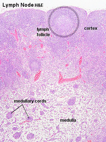
Lymph Node Histology
(Stain - Haematoxylin Eosin)
- Identify the connective tissue capsule and trabeculae, cortex and medulla of the lymph node, lymph nodules with germinal centres, medullary cords and postcapillary venules.
- Note the subcapsular and cortical sinus system was hardly (if at all) visible in these slides.
- Lymph Node Histology: Subcapsular Sinus | Follicle | Germinal Centre | Medullary Cords and Sinuses | High Endothelial Venules | Macrophages | Node cartoons
Links: Histology | Histology Stains | Blue Histology images copyright Lutz Slomianka 1998-2009. The literary and artistic works on the original Blue Histology website may be reproduced, adapted, published and distributed for non-commercial purposes. See also the page Histology Stains.
Cite this page: Hill, M.A. (2024, May 23) Embryology Lymph node histology 02.jpg. Retrieved from https://embryology.med.unsw.edu.au/embryology/index.php/File:Lymph_node_histology_02.jpg
- © Dr Mark Hill 2024, UNSW Embryology ISBN: 978 0 7334 2609 4 - UNSW CRICOS Provider Code No. 00098G
File history
Click on a date/time to view the file as it appeared at that time.
| Date/Time | Thumbnail | Dimensions | User | Comment | |
|---|---|---|---|---|---|
| current | 18:25, 25 February 2012 | | 450 × 600 (130 KB) | Z8600021 (talk | contribs) | |
| 09:00, 14 February 2011 |  | 300 × 400 (73 KB) | S8600021 (talk | contribs) | ==Lymph Node Histology== Original file name: Lyn041he.jpg http://www.lab.anhb.uwa.edu.au/mb140/CorePages/Lymphoid1/lymph1.htm#Lymph Lymph node histology 02.jpg {{Template:Blue Histology}} Category:Immune |
You cannot overwrite this file.